
This article needs additional citations for verification. Please help improve this article by adding citations to reliable sources. Unsourced material may be challenged and removed. Find sources: "Cochlear duct" – news · newspapers · books · scholar · JSTOR (January 2009) (Learn how and when to remove this message) |
| Cochlear duct | |
|---|---|
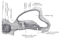
| 👁 Image Inner ear, with cochlear duct labeled near bottom. | |
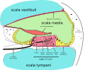
| 👁 Image Diagrammatic longitudinal section of the cochlea. (visible at far right under latin name ductus cochlearis) | |
| Details | |
| System | Ear |
| Identifiers | |
| Latin | ductus cochlearis |
| MeSH | D003053 |
| NeuroLex ID | birnlex_2562 |
| TA98 | A15.3.03.093 A15.3.03.092 |
| TA2 | 7022 |
| FMA | 79789 61119, 79789 |
| Anatomical terminology | |
The cochlear duct (a.k.a. the scala media) is an endolymph filled cavity inside the cochlea, located between the tympanic duct and the vestibular duct, separated by the basilar membrane and the vestibular membrane (Reissner's membrane) respectively. The cochlear duct houses the organ of Corti.[1]
Structure
[edit]The cochlear duct is part of the cochlea. It is separated from the tympanic duct (scala tympani) by the basilar membrane.[2] It is separated from the vestibular duct (scala vestibuli) by the vestibular membrane (Reissner's membrane).[2] The stria vascularis is located in the wall of the cochlear duct.[2]
Development
[edit]The cochlear duct develops from the ventral otic vesicle (otocyst).[3] It grows slightly flattened between the middle and outside of the body.[3] This development may be regulated by the genes EYA1, SIX1, GATA3, and TBX1.[3] The organ of Corti develops inside the cochlear duct.[4]
Function
[edit]The cochlear duct contains the organ of Corti.[2][5] This is attached to the basilar membrane.[5] It also contains endolymph, which contains high concentrations of K+ for the function of inner hair cells and outer hair cells in the organ of Corti.[2]
Clinical significance
[edit]Drugs delivered directly to the tympanic duct will spread to all of the cochlea except for the cochlear duct.[6] Rarely, the cochlear duct may develop to have the wrong shape.[3]
Additional images
[edit]-
Transverse section of the cochlear duct of a fetal cat.
-
The membranous labyrinth.
-
Floor of ductus cochlearis.
-
Cross section of the cochlea.
References
[edit]- ^ Hayes, Sarah H.; Ding, Dalian; Salvi, Richard J.; Allman, Brian L. (2013). "1 – Anatomy and physiology of the external, middle and inner ear". Handbook of Clinical Neurophysiology. Vol. 10. Elsevier. pp. 3–23. doi:10.1016/B978-0-7020-5310-8.00001-6. ISBN 978-0-7020-5310-8. ISSN 1567-4231.
- ^ a b c d e Feher, Joseph (2012). "4.7 – Hearing". Quantitative Human Physiology – An Introduction (2nd ed.). Academic Press. pp. 440–455. doi:10.1016/B978-0-12-800883-6.00040-9. ISBN 978-0-12-800883-6.
- ^ a b c d Fekete, D. M. (2009). "Cochlear Development". Encyclopedia of Neuroscience. Academic Press. pp. 1031–1040. doi:10.1016/B978-008045046-9.00252-7. ISBN 978-0-08-045046-9.
- ^ Goodyear, Richard J.; Richardson, Guy P. (2018). "6 – Structure, Function, and Development of the Tectorial Membrane: An Extracellular Matrix Essential for Hearing". Current Topics in Developmental Biology. Vol. 130. Elsevier. pp. 217–244. doi:10.1016/bs.ctdb.2018.02.006. ISBN 978-0-12-809802-8. ISSN 0070-2153. PMID 29853178.
- ^ a b Spelman, Francis A. (2013). "2.5.11 – Cochlear Prostheses". Biomaterials Science – An Introduction to Materials in Medicine (3rd ed.). Academic Press. pp. 967–980. doi:10.1016/B978-0-08-087780-8.00083-8. ISBN 978-0-12-374626-9.
- ^ Shepherd, R. K.; Seligman, P. M.; Fallon, J. B. (2015). "5 – Neuroprostheses for restoring hearing loss". Implantable Neuroprostheses for Restoring Function. Woodhead Publishing. pp. 97–125. doi:10.1016/B978-1-78242-101-6.00005-7. ISBN 978-1-78242-101-6.
External links
[edit]- Cross section at avatar.com.au