VOOZH about

URL: https://pubmed.ncbi.nlm.nih.gov/20029451/

⇱ Role of CCL2 (MCP-1) in traumatic brain injury (TBI): evidence from severe TBI patients and CCL2-/- mice - PubMed


Clipboard, Search History, and several other advanced features are temporarily unavailable.
Skip to main page content
πŸ‘ Dot gov

The .gov means it’s official.
Federal government websites often end in .gov or .mil. Before sharing sensitive information, make sure you’re on a federal government site.

πŸ‘ Https

The site is secure.
The https:// ensures that you are connecting to the official website and that any information you provide is encrypted and transmitted securely.

Access keys NCBI Homepage MyNCBI Homepage Main Content Main Navigation

Add to Collections

Add to My Bibliography

Your saved search

Create a file for external citation management software

Your RSS Feed

Abstract

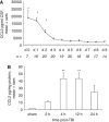
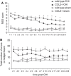
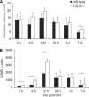
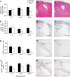
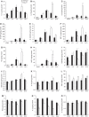
Cerebral inflammation involves molecular cascades contributing to progressive damage after traumatic brain injury (TBI). The chemokine CC ligand-2 (CCL2) (formerly monocyte chemoattractant protein-1, MCP-1) is implicated in macrophage recruitment into damaged parenchyma after TBI. This study analyzed the presence of CCL2 in human TBI, and further investigated the role of CCL2 in physiological and cellular mechanisms of secondary brain damage after TBI. Sustained elevation of CCL2 was detected in the cerebrospinal fluid (CSF) of severe TBI patients for 10 days after trauma, and in cortical homogenates of C57Bl/6 mice, peaking at 4 to 12 h after closed head injury (CHI). Neurological outcome, lesion volume, macrophage/microglia infiltration, astrogliosis, and the cerebral cytokine network were thus examined in CCL2-deficient (-/-) mice subjected to CHI. We found that CCL2-/- mice showed altered production of multiple cytokines acutely (2 to 24 h); however, this did not affect lesion size or cell death within the first week after CHI. In contrast, by 2 and 4 weeks, a delayed reduction in lesion volume, macrophage accumulation, and astrogliosis were observed in the injured cortex and ipsilateral thalamus of CCL2-/- mice, corresponding to improved functional recovery as compared with wild-type mice after CHI. Our findings confirm the significant role of CCL2 in mediating post-traumatic secondary brain damage.


Figures

πŸ‘ Figure 1
Figure 1
The time profiles of CCL2 production after TBI in humans and mice. CSF from severe TBI patients (n=21) was collected by drainage of the intraventricular catheter from day 0 (day of admission) to day 9, and CCL2 levels were measured by ELISA (A). A significant increase in CCL2 was evident on day 0, which remained elevated at day 1 and 2, as compared with subsequent times (*P<0.001). CCL2 levels were consistently higher in trauma patients as compared with those in control patients (dotted line; 717.01Β±71.5 pg/mL; P<0.001). CCL2 was also measured in the homogenates of lesioned cortex from wt mice (n=5) up to 24 h after CHI (B). CCL2 levels were significantly enhanced at 4 and 12 h after CHI as compared with those in sham-operated mice (**P<0.01). In both patients and mice, CCL2 levels peaked within the first 24 h. Data are expressed as mean+s.e.m.
πŸ‘ Figure 2
Figure 2
CCL2βˆ’/βˆ’ mice have improved functional recovery after CHI. The NSS reflects motor function, alertness, and behavior, with a score of 10 indicating the most severe dysfunction (A). CCL2βˆ’/βˆ’ mice (n=8) have similar scores at 1 h after injury as compared with wt mice (P=0.347, n=9), and show a comparable rate of recovery up to 6 days after CHI. However, CCL2βˆ’/βˆ’ mice showed improved neurological recovery, with significantly lower NSS scores compared with wt mice at 1, 7, 10, 14, 17, 21 and 24 days after CHI (*P<0.05, Tukey's post hoc). No differences were observed between sham-operated mice of each strain throughout the study period (P=0.863, n=4 to 5). The ledged beam test was also used to assess motor deficit of limbs contralateral to the lesioned cortex (B). The number of errors tended to be lower overall in CCL2βˆ’/βˆ’ mice as compared with that in wt mice after CHI (P=0.115). Data are expressed as mean+s.e.m.
πŸ‘ Figure 3
Figure 3
Lesion volume and cell death in wt and CCL2βˆ’/βˆ’ mice over the acute time course. No differences were detected in total lesion volume between wt and CCL2βˆ’/βˆ’ brains, as assessed using H&E-stained sections over 7 days after CHI (A; P=0.275, two-way ANOVA; n=6 to 7). By 7 days, lesion volume had decreased overall regardless of strain as compared with the lesion volume in the first 12 h after injury (*P<0.05; 12 h versus 7 days). Cell death was assessed by TUNEL staining using sections adjacent to H&E (B). Note the typical time course of cell death after TBI, with minimal positive cells at 2 and 4 h, followed by a dramatic peak of cell death at 12 h (P<0.05; 12 versus 2 h, 4 h, and 7 days). Consistent with lesion volume measurements, quantification of TUNEL labeling showed similar numbers of dead/dying cells in wt and CCL2βˆ’/βˆ’ brains (P=0.059, two-way ANOVA).
πŸ‘ Figure 4
Figure 4
Immunohistochemical analysis of the injured hemisphere in wt and CCL2βˆ’/βˆ’ mice over the delayed time course. Total lesion volumes in wt and CCL2βˆ’/βˆ’ mice was assessed using H&E-stained sections (A; n=6 to 7, from 7 to 28 days). Significantly reduced lesion volumes were observed in CCL2βˆ’/βˆ’ mice overall (P=0.047, two-way ANOVA), particularly at 28 days (*P<0.05, Tukey's post hoc). Representative wt and CCL2βˆ’/βˆ’ sections (B, C) show the evident difference in lesion size at 28 days. The loss of NeuN-positive neurons (D) coincided with changes in lesion volume, with a trend toward reduced neuronal loss in CCL2βˆ’/βˆ’ mice (F) at 28 days as compared with wt mice (E; P=0.088). Quantification of macrophages and reactive microglia was expressed as volume of F4/80-positive cell spread in the injured cortex (G). Despite similar volumes in both strains up to 7 days after CHI, by 14 and 28 days the volume of F4/80-positive cells was significantly reduced in CCL2βˆ’/βˆ’ mice (P=0.007 overall, two-way ANOVA; *P<0.05, Tukey's post hoc), as shown in representative sections of wt (H) and CCL2βˆ’/βˆ’ mice (I). Density of GFAP-positive astrocytes was calculated as the percentage of the cortical ROI (J). Across the time course, GFAP immunoreactivity in CCL2βˆ’/βˆ’ mice was reduced by approximately 30% as compared with that in wt mice (P=0.026, two-way ANOVA). This difference is evident in representative sections from wt (K) and CCL2βˆ’/βˆ’ mice at 28 days (L). Data are expressed as mean+s.e.m. Scale bar=1,000 μm in panels B, C, E, and F; 500 μm in panels H, I, K, and L. Color figure provided as Supplementary Information.
πŸ‘ Figure 5
Figure 5
Neuronal loss and glial activation in the ipsilateral thalamus of wt and CCL2βˆ’/βˆ’ mice. Neuronal loss in the ipsilateral dorsal thalamus was determined as % NeuN-positive staining of the uninjured contralateral thalamus (A; n=6 to 7). Overall, wt mice had a greater loss of NeuN staining as compared with CCL2βˆ’/βˆ’ mice (P=0.032, two-way ANOVA), as shown in representative sections (B, C; wt and CCL2βˆ’/βˆ’ mice at 28 days, respectively). The number of F4/80-positive cells was counted in the thalamus at 28 days (D). While abundant reactive microglia were observed in wt mice (E), CCL2βˆ’/βˆ’ mice (F) had significantly fewer F4/80-positive cells (*P<0.05, t-test; n=7). Astrocyte activation in the thalamus was expressed as the percentage of the ROI stained with GFAP (G). GFAP-staining intensity increased over time in both strains (**P<0.01, ***P<0.001, versus 7 days; two-way ANOVA effect of time). However, CCL2βˆ’/βˆ’ mice had overall less astrocyte reactivity as compared with that in wt mice (P=0.026). Representative images of the GFAP-stained ipsilateral thalamus are shown for wt (H) and CCL2βˆ’/βˆ’ (I) mice at 28 days. Data are expressed as mean+s.e.m. Scale bar=200 μm. Color figure provided as Supplementary Information.
πŸ‘ Figure 6
Figure 6
Localization of CCR2 receptor on F4/80-positive amoeboid macrophages. Double-labeling immunofluorescence of CCR2, in combination with NeuN, GFAP, or F4/80 illustrates the cellular localization of CCR2 at 4 days after CHI in wt mice. In the pericontusional cortex adjacent to the lesion core, no colocalization of NeuN or GFAP was detected with CCR2 staining (upper and middle panels). In contrast, CCR2 expression was evident within a subset of F4/80-positive amoeboid macrophages accumulating in the lesion (lower panels; arrows). Scale bar=100 μm.
πŸ‘ Figure 7
Figure 7
Inflammatory cytokine/chemokine expression in brain homogenates after CHI. The levels of 15 cytokines were detected in the homogenates of lesioned cortex over 24 h after CHI in wt and CCL2βˆ’/βˆ’ mice (n=5). Production of the cytokines/chemokines IL-1Ξ±, IL-1Ξ², IL-6, granulocyte colony-stimulating factor, IL-12(p40), MIP-1Ξ±, KC, and MIP-2 (A–H) was maximal and delayed to 12 h in CCL2βˆ’/βˆ’ mice (white bars) as compared with that in wt mice (black bars), in which they peaked by 4 h (*P<0.05, wt versus CCL2βˆ’/βˆ’ at 12 h, Tukey's post hoc). The chemokine RANTES (I) was reduced in CCL2βˆ’/βˆ’ mice as compared with wt mice, particularly in sham-operated mice and at 4 h after CHI (*P<0.05). Cytokines GM-CSF and IL-10 (J, K) were significantly elevated in CCL2βˆ’/βˆ’ (white bars) mice as compared with wt mice (black bars) at 12 h post CHI (*P<0.05). While TNFΞ± did not appear to be induced by CHI (L), overall the level of this cytokine was significantly higher in CCL2βˆ’/βˆ’ mice, even in sham-operated controls (P<0.001, two-way ANOVA). In contrast, the production of interferon-Ξ³, IL-2, and IL-12(p70) (M, N, and O) was reduced in CCL2βˆ’/βˆ’ mice as compared with that in wt controls over the time course (P=0.017, 0.001, and 0.033, respectively). Data are expressed as pg/mg protein (mean+s.e.m.).

References

    1. Banisadr G, Queraud-Lesaux F, Boutterin MC, Pelaprat D, Zalc B, Rostene W, Haour F, Parsadaniantz SM. Distribution, cellular localisation and functional role of CCR2 chemokine receptors in adult rat brain. J Neurochem. 2002;81:257–269. - PubMed
    1. Beni-Adani L, Gozes I, Cohen Y, Assaf Y, Steingart RA, Brenneman DE, Eizenberg O, Trembolver V, Shohami E. A peptide derived from activity-dependent neuroprotective protein (ADNP) ameliorates injury response in closed head injury in mice. J Pharmacol Exp Ther. 2001;296:57–63. - PubMed
    1. Berman JW, Guida MP, Warren J, Amat J, Brosnan CF. Localisation of monocyte chemoattractant peptide-1 expression in the central nervous system in experimental autoimmune encephalomyelitis and trauma in the rat. J Immunol. 1996;156:3017–3023. - PubMed
    1. Buttram SD, Wisniewski SR, Jackson EK, Adelson PD, Feldman K, Bayir H, Berger RP, Clark RS, Kochanek PM. Multiplex assessment of cytokine and chemokine levels in cerebrospinal fluid following severe pediatric traumatic brain injury: effects of moderate hypothermia. J Neurotrauma. 2007;24:1707–1717. - PubMed
    1. Bye N, Habgood MD, Callaway JK, Malakooti N, Potter A, Kossmann T, Morganti-Kossmann MC. Transient neuroprotection by minocycline following traumatic brain injury is associated with attenuated microglial activation but no changes in cell apoptosis or neutrophil infiltration. Exp Neurol. 2007;204:220–233. - PubMed
Cite

NCBI Literature Resources

MeSH PMC Bookshelf Disclaimer

The PubMed wordmark and PubMed logo are registered trademarks of the U.S. Department of Health and Human Services (HHS). Unauthorized use of these marks is strictly prohibited.