
Display options
Format
No abstract available
Figures
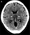
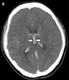
A, Image from noncontrast head CT demonstrates symmetric hypoattenuation within the bilateral medial thalami (arrows). B, Axial CT venogram demonstrates patency of the cerebral venous vasculature, including the internal cerebral veins (arrows). C, Coronal reformat of aCT angiogram demonstrates normal appearance of the basilar artery and proximal posterior cerebral arteries.
A, Image from noncontrast head CT demonstrates symmetric hypoattenuation within the bilateral medial thalami (arrows). B, Axial CT venogram demonstrates patency of the cerebral venous vasculature, including the internal cerebral veins (arrows). C, Coronal reformat of aCT angiogram demonstrates normal appearance of the basilar artery and proximal posterior cerebral arteries.
A, Image from noncontrast head CT demonstrates symmetric hypoattenuation within the bilateral medial thalami (arrows). B, Axial CT venogram demonstrates patency of the cerebral venous vasculature, including the internal cerebral veins (arrows). C, Coronal reformat of aCT angiogram demonstrates normal appearance of the basilar artery and proximal posterior cerebral arteries.
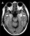
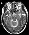
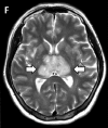
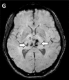
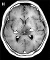
MRI images demonstrate T2 FLAIR hyperintensity within the bilateral medial temporal lobes and thalami (A, B, E, F) with evidence of hemorrhage indicated by hypointense signal intensity on susceptibility-weighted images (C, G) and rim enhancement on postcontrast images (D, H).
MRI images demonstrate T2 FLAIR hyperintensity within the bilateral medial temporal lobes and thalami (A, B, E, F) with evidence of hemorrhage indicated by hypointense signal intensity on susceptibility-weighted images (C, G) and rim enhancement on postcontrast images (D, H).
MRI images demonstrate T2 FLAIR hyperintensity within the bilateral medial temporal lobes and thalami (A, B, E, F) with evidence of hemorrhage indicated by hypointense signal intensity on susceptibility-weighted images (C, G) and rim enhancement on postcontrast images (D, H).
MRI images demonstrate T2 FLAIR hyperintensity within the bilateral medial temporal lobes and thalami (A, B, E, F) with evidence of hemorrhage indicated by hypointense signal intensity on susceptibility-weighted images (C, G) and rim enhancement on postcontrast images (D, H).
MRI images demonstrate T2 FLAIR hyperintensity within the bilateral medial temporal lobes and thalami (A, B, E, F) with evidence of hemorrhage indicated by hypointense signal intensity on susceptibility-weighted images (C, G) and rim enhancement on postcontrast images (D, H).
MRI images demonstrate T2 FLAIR hyperintensity within the bilateral medial temporal lobes and thalami (A, B, E, F) with evidence of hemorrhage indicated by hypointense signal intensity on susceptibility-weighted images (C, G) and rim enhancement on postcontrast images (D, H).
MRI images demonstrate T2 FLAIR hyperintensity within the bilateral medial temporal lobes and thalami (A, B, E, F) with evidence of hemorrhage indicated by hypointense signal intensity on susceptibility-weighted images (C, G) and rim enhancement on postcontrast images (D, H).
MRI images demonstrate T2 FLAIR hyperintensity within the bilateral medial temporal lobes and thalami (A, B, E, F) with evidence of hemorrhage indicated by hypointense signal intensity on susceptibility-weighted images (C, G) and rim enhancement on postcontrast images (D, H).
Comment in
-
New-onset psychosis in COVID-19 pandemic: a case series in Madrid.Rentero D, Juanes A, Losada CP, Álvarez S, Parra A, Santana V, Martí I, Urricelqui J. Rentero D, et al. Psychiatry Res. 2020 Aug;290:113097. doi: 10.1016/j.psychres.2020.113097. Epub 2020 May 13. Psychiatry Res. 2020. PMID: 32480119 Free PMC article. No abstract available.
-
Clinical Course of a Patient with Radiographically Described Acute Necrotizing Encephalopathy.Bansal P, Fory EK, Malik S, Memon AB. Bansal P, et al. Radiology. 2020 Nov;297(2):E278-E280. doi: 10.1148/radiol.2020203132. Epub 2020 Aug 13. Radiology. 2020. PMID: 32787703 Free PMC article. No abstract available.
References
-
- Coronavirus disease (COVID-19) Pandemic . Geneva: World Health Organization, March 23, 2020 (https://www.who.int/emergencies/diseases/novel-coronavirus-2019).
-
- Rossi A. Imaging of acute disseminated encephalomyelitis. Neuroimaging Clinics, 18(1): 149-161. - PubMed
Publication types
MeSH terms
LinkOut - more resources
Full Text Sources
Other Literature Sources
Medical