
Display options
Format
No abstract available
Figures
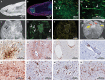
Panel A (magnetic resonance microscopy of the olfactory bulb) shows an area of hyperintense signal (arrow). Panel B shows the corresponding area on multiplex immunofluorescence imaging, which revealed a focal area of fibrinogen leakage (in the box, fibrinogen is shown in green, collagen IV is shown in yellow, and nuclei are shown in blue). Panel B1 shows diffuse leakage of fibrinogen in the parenchyma (an enlarged view showing marked blood vessel staining for collagen IV is shown in Panel B2). Panel B2 (collagen IV immunostaining) shows intact (arrowhead) and thinned (arrow) basal lamina with fibrinogen leakage into the parenchyma. Panel C shows magnetic resonance microscopy of the pons, and Panel D (fibrinogen staining) shows areas of increased signal intensity corresponding to the vascular leakage visible on magnetic resonance microscopy. The arrows and the area within the dashed lines in Panels C and D indicate the vascular leakage. Panels A through E represent imaging performed in Patient IA1. Panel E (collagen IV immunostaining) shows areas of fibrinogen leakage in blood vessels in Patient IA1. Panel F shows magnetic resonance microscopy of the medulla in Patient IA3. The yellow arrows indicate linear hypointense signals, and the red arrows indicate linear hyperintense signals. Panel G shows CD68+ perivascular macrophages in the pons in Patient NY6. Panel H shows perivascular astrocytosis in the basal ganglia in Patient NY5. Panel I shows perivascular CD3+ cells in the cerebellum in Patient IA1. Panel J shows intraluminal and perivascular CD8+ cells in the pons in Patient NY6. Panel K shows perineuronal IBA1 cells in the pons in Patient NY6. Panel L shows CD68+ cells in the dorsal motor nucleus of the vagus nerve in Patient IA1. Panel M shows a solitary nucleus in the medulla and Panel N shows a pre-Bötzinger complex in Patient IA1. (Diaminobenzidine staining was used in Panels G through N.)
Comment in
-
Virtual Biopsy: A Reality Thanks to Advances in Radiology.Aragao MFVV, Oliveira ADP, Lima ARMC, Leal MC, Valenca MM. Aragao MFVV, et al. AJNR Am J Neuroradiol. 2021 Jul;42(7):E37-E38. doi: 10.3174/ajnr.A7092. Epub 2021 Mar 25. AJNR Am J Neuroradiol. 2021. PMID: 33766826 Free PMC article. No abstract available.
-
Post-COVID-19 neuropsychiatric syndrome: Is maladaptive glial recovery to blame?Steardo L Jr, Steardo L, Verkhratsky A, Scuderi C. Steardo L Jr, et al. Acta Physiol (Oxf). 2021 Oct;233(2):e13717. doi: 10.1111/apha.13717. Epub 2021 Jul 22. Acta Physiol (Oxf). 2021. PMID: 34264006 Free PMC article. No abstract available.
References
Publication types
MeSH terms
Grants and funding
LinkOut - more resources
Full Text Sources
Other Literature Sources
Medical