
1MVC | pdb_00001mvc
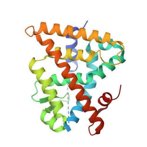
Crystal structure of the human RXR alpha ligand binding domain bound to the synthetic agonist compound BMS 649 and a coactivator peptide
- PDB DOI: https://doi.org/10.2210/pdb1MVC/pdb
- Classification: TRANSCRIPTION
- Organism(s): Homo sapiens
- Expression System: Escherichia coli BL21(DE3)
- Mutation(s): No
- Deposited: 2002-09-24 Released: 2002-10-16
- Deposition Author(s): Egea, P.F., Mitschler, A., Moras, D.
Experimental Data Snapshot
- Method: X-RAY DIFFRACTION
- Resolution: 1.90 Å
- R-Value Free: 0.228 (Depositor)
- R-Value Work: 0.201 (Depositor)
- R-Value Observed: 0.201 (Depositor)
Starting Model: experimental
View more details
wwPDB Validation 3D Report Full Report
- 👁 Image
Download Mendeley
Molecular Recognition of Agonist Ligands by RXRs
Egea, P.F., Mitschler, A., Moras, D.(2002) Mol Endocrinol 16: 987-997
- PubMed: 11981034 Search on PubMed
- DOI: https://doi.org/10.1210/mend.16.5.0823
- Primary Citation Related Structures:
1MV9, 1MVC, 1MZN - PubMed Abstract:
The nuclear receptor RXR is an obligate partner in many signal transduction pathways. We report the high-resolution structures of two complexes of the human RXRalpha ligand-binding domain specifically bound to two different and chemically unrelated agonist compounds: docosa hexaenoic acid, a natural derivative of eicosanoic acid, present in mammalian cells and recently identified as a potential endogenous RXR ligand in the mouse brain, and the synthetic ligand BMS 649. In both structures the RXR-ligand-binding domain forms homodimers and exhibits the active conformation previously observed with 9-cis-RA. Analysis of the differences in ligand-protein contacts (predominantly van der Waals forces) and binding cavity geometries and volumes for the several agonist-bound RXR structures clarifies the structural features important for ligand recognition. The L-shaped ligand-binding pocket adapts to the diverse ligands, especially at the level of residue N306, which might thus constitute a new target for drug-design. Despite its highest affinity 9-cis-RA displays the lowest number of ligand-protein contacts. These structural results support the idea that docosa hexaenoic acid and related fatty acids could be natural agonists of RXRs and question the real nature of the endogenous ligand(s) in mammalian cells.
- Laboratoire de Biologie et Génomique Structurales, Université Louis Pasteur, Parc d'Innovation BP163, 67404 Illkirch cedex, France.
Organizational Affiliation:
Explore in 3D: Structure | Sequence Annotations | Validation Report | Ligand Interaction (BM6)
Explore in 3D: Structure | Sequence Annotations | Validation Report | Ligand Interaction (BM6)
Global Symmetry: Cyclic - C2 (Explore in 3D)
Global Stoichiometry: Hetero 4-mer - A2B2
Pseudo Symmetry: Asymmetric - C1
Pseudo Stoichiometry: Hetero 4-mer - A2B1C1
Find Similar Assemblies
Biological assembly 1 assigned by authors.
Macromolecule Content
- Total Structure Weight: 28.82 kDa
- Atom Count: 1,988
- Modeled Residue Count: 223
- Deposited Residue Count: 253
- Unique protein chains: 2
Entity ID: 1 | |||||
|---|---|---|---|---|---|
| Molecule | Chains | Sequence Length | Organism | Details | Image |
| RXR retinoid X receptor | 240 | Homo sapiens | Mutation(s): 0 | 👁 Image | |
UniProt & NIH Common Fund Data Resources | |||||
Find proteins for P19793 (Homo sapiens) Explore P19793 Go to UniProtKB: P19793 | |||||
PHAROS: P19793 GTEx: ENSG00000186350 | |||||
Entity Groups | |||||
| Sequence Clusters | 30% Identity50% Identity70% Identity90% Identity95% Identity100% Identity | ||||
| UniProt Group | P19793 | ||||
Sequence AnnotationsExpand | |||||
| |||||
Find similar proteins by: Sequence | 3D Structure
Entity ID: 2 | |||||
|---|---|---|---|---|---|
| Molecule | Chains | Sequence Length | Organism | Details | Image |
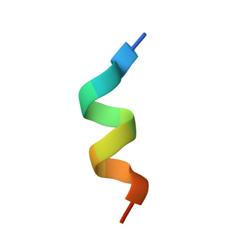
| Nuclear receptor coactivator 2 | 13 | N/A | Mutation(s): 0 | 👁 Image | |
UniProt & NIH Common Fund Data Resources | |||||
Find proteins for Q15596 (Homo sapiens) Explore Q15596 Go to UniProtKB: Q15596 | |||||
PHAROS: Q15596 GTEx: ENSG00000140396 | |||||
Entity Groups | |||||
| Sequence Clusters | 30% Identity50% Identity70% Identity90% Identity95% Identity100% Identity | ||||
| UniProt Group | Q15596 | ||||
Sequence AnnotationsExpand | |||||
| |||||
| Ligands 1 Unique | |||||
|---|---|---|---|---|---|
| ID | Chains | Name / Formula / InChI Key | 2D Diagram | 3D Interactions | |
| BM6 Query on BM6 Download Ideal Coordinates CCD File | C [auth A] | 4-[2-(5,5,8,8-TETRAMETHYL-5,6,7,8-TETRAHYDRO-NAPHTHALEN-2-YL)-[1,3]DIOXOLAN-2-YL]-BENZOIC ACID C24 H27 O4 ZZUKALQMHNSWTK-UHFFFAOYSA-M | 👁 Image | ||
Experimental Data
- Method: X-RAY DIFFRACTION
- Resolution: 1.90 Å
- R-Value Free: 0.228 (Depositor)
- R-Value Work: 0.201 (Depositor)
- R-Value Observed: 0.201 (Depositor)
| Length ( Å ) | Angle ( ˚ ) |
|---|---|
| a = 64.511 | α = 90 |
| b = 64.511 | β = 90 |
| c = 111.465 | γ = 90 |
| Software Name | Purpose |
|---|---|
| DENZO | data reduction |
| SCALEPACK | data scaling |
| AMoRE | phasing |
| CNS | refinement |
Deposition Data
- Released Date: 2002-10-16 Deposition Author(s): Egea, P.F., Mitschler, A., Moras, D.
Revision History (Full details and data files)
- Version 1.0: 2002-10-16
Type: Initial release - Version 1.1: 2008-04-28
Changes: Version format compliance - Version 1.2: 2011-07-13
Changes: Version format compliance - Version 1.3: 2023-10-25
Changes: Data collection, Database references, Derived calculations, Refinement description