
- FASTA Sequence
- PDBx/mmCIF Format
- PDBx/mmCIF Format (gz)
- BinaryCIF Format (gz)
- Legacy PDB Format
- Legacy PDB Format (gz)
- PDBML/XML Format (gz)
- NMR Restraints (Text)
- NMR Restraints (Text - gz)
- NMR Restraints v2 (Text)
- NMR Restraints v2 (Text - gz)
- Validation Full (PDF - gz)
- Validation (XML - gz)
- Validation (CIF - gz)
- Biological Assembly 1 (CIF - gz)
- Biological Assembly 1 (PDB - gz)
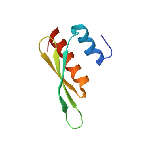
2B7T | pdb_00002b7t
Structure of ADAR2 dsRBM1
- PDB DOI: https://doi.org/10.2210/pdb2B7T/pdb
- Classification: HYDROLASE
- Organism(s): Rattus norvegicus
- Expression System: Escherichia coli BL21(DE3)
- Mutation(s): No
- Deposited: 2005-10-05 Released: 2006-03-14
- Deposition Author(s): Stefl, R., Xu, M., Skrisovska, L., Emeson, R.B., Allain, F.H.-T.
Experimental Data Snapshot
- Method: SOLUTION NMR
- Conformers Calculated: 50
- Conformers Submitted: 20
- Selection Criteria: target function
wwPDB Validation3D Report Full Report
Literature
- 👁 Image
Download Mendeley
Structure and specific RNA binding of ADAR2 double-stranded RNA binding motifs.
Stefl, R., Xu, M., Skrisovska, L., Emeson, R.B., Allain, F.H.-T.(2006) Structure 14: 345-355
- PubMed: 16472753 Search on PubMed
- DOI: https://doi.org/10.1016/j.str.2005.11.013
- Primary Citation Related Structures:
2B7T, 2B7V - PubMed Abstract:
Adenosine deaminases that act on RNA (ADARs) site-selectively modify adenosines to inosines within RNA transcripts, thereby recoding genomic information. How ADARs select specific adenosine moieties for deamination is poorly understood. Here, we report NMR structures of the two double-stranded RNA binding motifs (dsRBMs) of rat ADAR2 and an NMR chemical shift perturbation study of the interaction of the two dsRBMs with a 71 nucleotide RNA encoding the R/G site of the GluR-B. We have identified the protein and the RNA surfaces involved in complex formation, allowing us to present an NMR-based model of the complex. We have found that dsRBM1 recognizes a conserved pentaloop, whereas dsRBM2 recognizes two bulged bases adjacent to the editing site, demonstrating RNA structure-dependent recognition by the ADAR2 dsRBMs. In vitro mutagenesis studies with both the protein and the RNA further support our structural findings.
- Institute of Molecular Biology and Biophysics, ETH Zürich, 8093 Zürich, Switzerland.
Organizational Affiliation:
Biological Assembly 1
Explore in 3D: Structure | Sequence Annotations | Validation Report
Global Symmetry: Asymmetric - C1
Global Stoichiometry: Monomer - A1
Find Similar Assemblies
Biological assembly 1 assigned by authors.
Macromolecule Content
- Total Structure Weight: 7.92 kDa
- Atom Count: 557
- Modeled Residue Count: 73
- Deposited Residue Count: 73
- Unique protein chains: 1
Macromolecules
Entity ID: 1 | |||||
|---|---|---|---|---|---|
| Molecule | Chains | Sequence Length | Organism | Details | Image |
| Double-stranded RNA-specific editase 1 | 73 | Rattus norvegicus | Mutation(s): 0 Gene Names: Adarb1, Red1 EC: 3.5 (PDB Primary Data), 3.5.4.37 (UniProt) | 👁 Image | |
UniProt | |||||
Entity Groups | |||||
| Sequence Clusters | 30% Identity50% Identity70% Identity90% Identity95% Identity100% Identity | ||||
| UniProt Group | P51400 | ||||
Sequence AnnotationsExpand | |||||
Reference Sequence | |||||
Experimental Data & Validation
Experimental Data
- Method: SOLUTION NMR
- Conformers Calculated: 50
- Conformers Submitted: 20
- Selection Criteria: target function
Entry History
Deposition Data
- Released Date: 2006-03-14 Deposition Author(s): Stefl, R., Xu, M., Skrisovska, L., Emeson, R.B., Allain, F.H.-T.
Revision History (Full details and data files)
- Version 1.0: 2006-03-14
Type: Initial release - Version 1.1: 2008-05-01
Changes: Version format compliance - Version 1.2: 2011-07-13
Changes: Version format compliance - Version 1.3: 2022-03-09
Changes: Data collection, Database references, Derived calculations - Version 1.4: 2024-05-22
Changes: Data collection