
- FASTA Sequence
- PDBx/mmCIF Format
- PDBx/mmCIF Format (gz)
- BinaryCIF Format (gz)
- Legacy PDB Format
- Legacy PDB Format (gz)
- PDBML/XML Format (gz)
- Structure Factors (CIF)
- Structure Factors (CIF - gz)
- Validation Full (PDF - gz)
- Validation (XML - gz)
- Validation (CIF - gz)
- Validation 2fo-fc coefficients (CIF - gz)
- Validation fo-fc coefficients (CIF - gz)
- Biological Assembly 1 (CIF - gz)
- Biological Assembly 2 (CIF - gz)
- Biological Assembly 3 (CIF - gz)
- Biological Assembly 4 (CIF - gz)
- Biological Assembly 5 (CIF - gz)
- Biological Assembly 6 (CIF - gz)
- Biological Assembly 1 (PDB - gz)
- Biological Assembly 2 (PDB - gz)
- Biological Assembly 3 (PDB - gz)
- Biological Assembly 4 (PDB - gz)
- Biological Assembly 5 (PDB - gz)
- Biological Assembly 6 (PDB - gz)
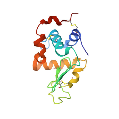
2G4N | pdb_00002g4n
Anomalous substructure of alpha-lactalbumin
- PDB DOI: https://doi.org/10.2210/pdb2G4N/pdb
- Classification: ALLERGEN
- Organism(s): Bos taurus
- Mutation(s): No
- Deposited: 2006-02-22 Released: 2007-02-20
- Deposition Author(s): Mueller-Dieckmann, C., Weiss, M.S.
Experimental Data Snapshot
- Method: X-RAY DIFFRACTION
- Resolution: 2.30 Å
- R-Value Free: 0.359 (Depositor)
- R-Value Work: 0.296 (Depositor), 0.294 (DCC)
- R-Value Observed: 0.297 (Depositor)
wwPDB Validation 3D Report Full Report
- 👁 Image
Download Mendeley
On the routine use of soft X-rays in macromolecular crystallography. Part IV. Efficient determination of anomalous substructures in biomacromolecules using longer X-ray wavelengths.
Mueller-Dieckmann, C., Panjikar, S., Schmidt, A., Mueller, S., Kuper, J., Geerlof, A., Wilmanns, M., Singh, R.K., Tucker, P.A., Weiss, M.S.(2007) Acta Crystallogr D Biol Crystallogr 63: 366-380
- PubMed: 17327674 Search on PubMed
- DOI: https://doi.org/10.1107/S0907444906055624
- Primary Citation Related Structures:
2G4H, 2G4I, 2G4J, 2G4K, 2G4L, 2G4M, 2G4N, 2G4O, 2G4P, 2G4Q, 2G4R, 2G4S, 2G4T, 2G4U, 2G4V, 2G4W, 2G4X, 2G4Y, 2G4Z, 2G51, 2G52, 2G55, 2ILL - PubMed Abstract:
23 different crystal forms of 19 different biological macromolecules were examined with respect to their anomalously scattering substructures using diffraction data collected at a wavelength of 2.0 A (6.2 keV). In more than 90% of the cases the substructure was found to contain more than just the protein S atoms. The data presented suggest that chloride, sulfate, phosphate or metal ions from the buffer or even from the purification protocol are frequently bound to the protein molecule and that these ions are often overlooked, especially if they are not bound at full occupancy. Thus, in order to fully describe the macromolecule under study, it seems desirable that any structure determination be complemented with a long-wavelength data set.
- EMBL Hamburg Outstation, c/o DESY, Notkestrasse 85, D-22603 Hamburg, Germany.
Organizational Affiliation:
Explore in 3D: Structure | Sequence Annotations | Electron Density | Validation Report | Ligand Interaction (CA)
Explore in 3D: Structure | Sequence Annotations | Electron Density | Validation Report | Ligand Interaction (CA)
Global Symmetry: Asymmetric - C1
Global Stoichiometry: Monomer - A1
Find Similar Assemblies
Biological assembly 1 assigned by authors.
Explore in 3D: Structure | Sequence Annotations | Electron Density | Validation Report | Ligand Interaction (CA)
Global Symmetry: Asymmetric - C1
Global Stoichiometry: Monomer - A1
Find Similar Assemblies
Biological assembly 2 assigned by authors.
Explore in 3D: Structure | Sequence Annotations | Electron Density | Validation Report | Ligand Interaction (CA)
Global Symmetry: Asymmetric - C1
Global Stoichiometry: Monomer - A1
Find Similar Assemblies
Biological assembly 3 assigned by authors.
Explore in 3D: Structure | Sequence Annotations | Electron Density | Validation Report | Ligand Interaction (CA)
Global Symmetry: Asymmetric - C1
Global Stoichiometry: Monomer - A1
Find Similar Assemblies
Biological assembly 4 assigned by authors.
Explore in 3D: Structure | Sequence Annotations | Electron Density | Validation Report | Ligand Interaction (CA)
Global Symmetry: Asymmetric - C1
Global Stoichiometry: Monomer - A1
Find Similar Assemblies
Biological assembly 5 assigned by authors.
Explore in 3D: Structure | Sequence Annotations | Electron Density | Validation Report | Ligand Interaction (CA)
Global Symmetry: Asymmetric - C1
Global Stoichiometry: Monomer - A1
Find Similar Assemblies
Biological assembly 6 assigned by authors.
Macromolecule Content
- Total Structure Weight: 85.53 kDa
- Atom Count: 5,938
- Modeled Residue Count: 732
- Deposited Residue Count: 738
- Unique protein chains: 1
Entity ID: 1 | |||||
|---|---|---|---|---|---|
| Molecule | Chains | Sequence Length | Organism | Details | Image |
| Alpha-lactalbumin | 123 | Bos taurus | Mutation(s): 0 | 👁 Image | |
UniProt | |||||
Find proteins for P00711 (Bos taurus) Explore P00711 Go to UniProtKB: P00711 | |||||
Entity Groups | |||||
| Sequence Clusters | 30% Identity50% Identity70% Identity90% Identity95% Identity100% Identity | ||||
| UniProt Group | P00711 | ||||
Sequence AnnotationsExpand | |||||
| |||||
| Ligands 2 Unique | |||||
|---|---|---|---|---|---|
| ID | Chains | Name / Formula / InChI Key | 2D Diagram | 3D Interactions | |
| CA Query on CA Download Ideal Coordinates CCD File
| G [auth A] I [auth B] K [auth C] L [auth D] M [auth E] G [auth A], I [auth B], K [auth C], L [auth D], M [auth E], N [auth F] | CALCIUM ION Ca BHPQYMZQTOCNFJ-UHFFFAOYSA-N | 👁 Image | ||
| K Query on K Download Ideal Coordinates CCD File | H [auth A], J [auth B] | POTASSIUM ION K NPYPAHLBTDXSSS-UHFFFAOYSA-N | 👁 Image | ||
Experimental Data
- Method: X-RAY DIFFRACTION
- Resolution: 2.30 Å
- R-Value Free: 0.359 (Depositor)
- R-Value Work: 0.296 (Depositor), 0.294 (DCC)
- R-Value Observed: 0.297 (Depositor)
| Length ( Å ) | Angle ( ˚ ) |
|---|---|
| a = 68.43 | α = 90 |
| b = 104.8 | β = 90 |
| c = 119.11 | γ = 90 |
| Software Name | Purpose |
|---|---|
| REFMAC | refinement |
| DENZO | data reduction |
| CCP4 | data scaling |
| FFT | phasing |
Deposition Data
- Released Date: 2007-02-20 Deposition Author(s): Mueller-Dieckmann, C., Weiss, M.S.
Revision History (Full details and data files)
- Version 1.0: 2007-02-20
Type: Initial release - Version 1.1: 2008-05-01
Changes: Version format compliance - Version 1.2: 2011-07-13
Changes: Advisory, Version format compliance - Version 1.3: 2024-11-13
Changes: Data collection, Database references, Derived calculations, Refinement description, Structure summary