
- FASTA Sequence
- PDBx/mmCIF Format
- PDBx/mmCIF Format (gz)
- BinaryCIF Format (gz)
- Legacy PDB Format
- Legacy PDB Format (gz)
- PDBML/XML Format (gz)
- Structure Factors (CIF)
- Structure Factors (CIF - gz)
- Validation Full (PDF - gz)
- Validation (XML - gz)
- Validation (CIF - gz)
- Validation 2fo-fc coefficients (CIF - gz)
- Validation fo-fc coefficients (CIF - gz)
- Biological Assembly 1 (CIF - gz)
- Biological Assembly 1 (PDB - gz)
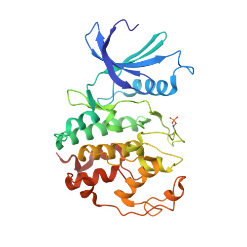
2JGZ | pdb_00002jgz
Crystal structure of phospho-CDK2 in complex with Cyclin B
- PDB DOI: https://doi.org/10.2210/pdb2JGZ/pdb
- Classification: TRANSFERASE
- Organism(s): Homo sapiens
- Expression System: Escherichia coli
- Mutation(s): No
- Deposited: 2007-02-17 Released: 2007-05-22
- Deposition Author(s): Brown, N.R., Petri, E., Lowe, E.D., Skamnaki, V., Johnson, L.N.
Experimental Data Snapshot
- Method: X-RAY DIFFRACTION
- Resolution: 2.90 Å
- R-Value Free: 0.275 (Depositor), 0.270 (DCC)
- R-Value Work: 0.205 (Depositor), 0.200 (DCC)
- R-Value Observed: 0.208 (Depositor)
Starting Model: experimental
View more details
wwPDB Validation 3D Report Full Report
- 👁 Image
Download Mendeley
Cyclin B and cyclin A confer different substrate recognition properties on CDK2.
Brown, N.R., Lowe, E.D., Petri, E., Skamnaki, V., Antrobus, R., Johnson, L.N.(2007) Cell Cycle 6: 1350-1359
- PubMed: 17495531 Search on PubMed
- DOI: https://doi.org/10.4161/cc.6.11.4278
- Primary Citation Related Structures:
2JGZ - PubMed Abstract:
The transitions of the cell cycle are regulated by the cyclin dependent protein kinases (CDKs). The cyclins activate their respective CDKs and confer substrate recognition properties. We report the structure of phospho-CDK2/cyclin B and show that cyclin B confers M phase-like properties on CDK2, the kinase that is usually associated with S phase. Cyclin B produces an almost identical activated conformation of CDK2 as that produced by cyclin A. There are differences between cyclin A and cyclin B at the recruitment site, which in cyclin A is used to recruit substrates containing an RXL motif. Because of sequence differences this site in cyclin B binds RXL motifs more weakly than in cyclin A. Despite similarity in kinase structures, phospho-CDK2/cyclin B phosphorylates substrates, such as nuclear lamin and a model peptide derived from p107, at sequences SPXX that differ from the canonical CDK2/cyclin A substrate recognition motif, SPXK. CDK2/cyclin B phosphorylation at these non-canonical sites is not dependent on the presence of a RXL recruitment motif. The p107 peptide contains two SP motifs each followed by a non-canonical sequence of which only one site (Ser640) is phosphorylated by pCDK2/cyclin A while two sites are phosphorylated by pCDK2/cyclin B. The second site is too close to the RXL motif to allow the cyclin A recruitment site to be effective, as previous work has shown that there must be at least 16 residues between the catalytic site serine and the RXL motif. Thus the cyclins A and B in addition to their role in promoting the activatory conformational switch in CDK2, also provide differential substrate specificity.
- Laboratory of Molecular Biophysics, Department of Biochemistry, University of Oxford, Oxford, UK.
Organizational Affiliation:
Explore in 3D: Structure | Sequence Annotations | Electron Density | Validation Report
Explore in 3D: Structure | Sequence Annotations | Electron Density | Validation Report
Global Symmetry: Asymmetric - C1
Global Stoichiometry: Hetero 2-mer - A1B1
Find Similar Assemblies
Biological assembly 1 assigned by authors and generated by PQS (software)
Macromolecule Content
- Total Structure Weight: 63.11 kDa
- Atom Count: 4,437
- Modeled Residue Count: 549
- Deposited Residue Count: 549
- Unique protein chains: 2
Entity ID: 1 | |||||
|---|---|---|---|---|---|
| Molecule | Chains | Sequence Length | Organism | Details | Image |
| CELL DIVISION PROTEIN KINASE 2 | 289 | Homo sapiens | Mutation(s): 0 EC: 2.7.1 (PDB Primary Data), 2.7.11.22 (UniProt) | 👁 Image | |
UniProt & NIH Common Fund Data Resources | |||||
Find proteins for P24941 (Homo sapiens) Explore P24941 Go to UniProtKB: P24941 | |||||
PHAROS: P24941 GTEx: ENSG00000123374 | |||||
Entity Groups | |||||
| Sequence Clusters | 30% Identity50% Identity70% Identity90% Identity95% Identity100% Identity | ||||
| UniProt Group | P24941 | ||||
Sequence AnnotationsExpand | |||||
| |||||
Entity ID: 2 | |||||
|---|---|---|---|---|---|
| Molecule | Chains | Sequence Length | Organism | Details | Image |
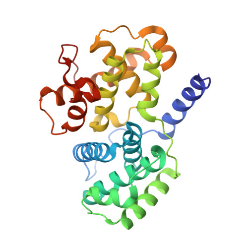
| G2/MITOTIC-SPECIFIC CYCLIN-B1 | 260 | Homo sapiens | Mutation(s): 0 | 👁 Image | |
UniProt & NIH Common Fund Data Resources | |||||
Find proteins for P14635 (Homo sapiens) Explore P14635 Go to UniProtKB: P14635 | |||||
PHAROS: P14635 GTEx: ENSG00000134057 | |||||
Entity Groups | |||||
| Sequence Clusters | 30% Identity50% Identity70% Identity90% Identity95% Identity100% Identity | ||||
| UniProt Group | P14635 | ||||
Sequence AnnotationsExpand | |||||
| |||||
| Modified Residues 1 Unique | |||||
|---|---|---|---|---|---|
| ID | Chains | Type | Formula | 2D Diagram | Parent |
| TPO Query on TPO | A | L-PEPTIDE LINKING | C4 H10 N O6 P | 👁 Image | THR |
Experimental Data
- Method: X-RAY DIFFRACTION
- Resolution: 2.90 Å
- R-Value Free: 0.275 (Depositor), 0.270 (DCC)
- R-Value Work: 0.205 (Depositor), 0.200 (DCC)
- R-Value Observed: 0.208 (Depositor)
| Length ( Å ) | Angle ( ˚ ) |
|---|---|
| a = 104.78 | α = 90 |
| b = 104.78 | β = 90 |
| c = 251.635 | γ = 120 |
| Software Name | Purpose |
|---|---|
| REFMAC | refinement |
| MOSFLM | data reduction |
| SCALA | data scaling |
| PHASER | phasing |
Deposition Data
- Released Date: 2007-05-22 Deposition Author(s): Brown, N.R., Petri, E., Lowe, E.D., Skamnaki, V., Johnson, L.N.
Revision History (Full details and data files)
- Version 1.0: 2007-05-22
Type: Initial release - Version 1.1: 2011-05-08
Changes: Version format compliance - Version 1.2: 2011-07-13
Changes: Version format compliance - Version 1.3: 2019-10-09
Changes: Data collection, Database references, Derived calculations, Other - Version 1.4: 2023-12-13
Changes: Data collection, Database references, Refinement description - Version 1.5: 2024-10-23
Changes: Structure summary