 |
VOOZH | about |
|
VOOZH | about |
Osteoporosis, in which low bone mass and micro-structural deterioration of bone tissue lead to increased bone fragility, is the most common metabolic bone disease in the United States. [1, 2] Osteoporosis can result in devastating physical, psychosocial, and economic consequences. Still, it is often overlooked and undertreated, in large part because it is clinically silent; there are no symptoms before a fracture occurs. (See the image below.)
Osteoporosis does not become clinically apparent until a fracture occurs and so is sometimes referred to as the “silent disease.” Two-thirds of vertebral fractures are painless, although patients may complain of the resulting stooped posture and height loss. Typical findings in patients with painful vertebral fractures may include the following:
Patients who have sustained a hip fracture may experience the following:
On physical examination, patients with vertebral compression fractures may demonstrate the following:
Patients with hip fractures may demonstrate the following:
Patients with Colles fractures may have the following:
Patients with pubic and sacral fractures may have the following:
Balance difficulties may be evident, especially in patients with an altered center of gravity from severe kyphosis. [3] Patients may have difficulty performing tandem gait and performing single-limb stance.
See the Presentation for more detail.
Baseline laboratory studies include the following:
Bone mineral density (BMD) measurement is recommended in the following patients [4] :
Dual-energy x-ray absorptiometry (DXA) is currently the criterion standard for the evaluation of BMD. [5, 6] Peripheral DXA is used to measure BMD at the wrist; it may be most useful in identifying patients at very low fracture risk who require no further workup.
DXA provides the patient’s T-score, which is the BMD value compared with that of control subjects who are at their peak BMD. [7, 8, 9] World Health Organization (WHO) criteria define a normal T-score value as within 1 standard deviation (SD) of the mean BMD value in a healthy young adult. Values lying farther from the mean are stratified as follows [8] :
DXA also provides the patient’s Z-score, which reflects a value compared with that of persons matched for age and sex. Z-scores adjusted for ethnicity or race should be used in the following patients:
Z-score values of –2.0 SD or lower are defined as "below the expected range for age" and those above –2.0 SD as "within the expected range for age." The diagnosis of osteoporosis in these groups should not be based on densitometric criteria alone.
The BHOF recommends considering imaging to detect subclinical vertebral fractures for the following patients [10] :
In addition, BHOF guidelines recommend vertebral imaging in postmenopausal women and men age ≥ 50 years with any of the following specific risk factors:
If bone density testing is not available, vertebral imaging may be considered based on age alone.
Other plain radiography features and recommendations are as follows:
See Workup for more detail.
Lifestyle modification for the prevention of osteoporotic fractures includes the following [11] :
Pharmacologic therapy
The NOF recommends reserving pharmacologic therapy for postmenopausal women and men aged 50 years or older who present with the following [4] :
Guidelines from the American Association of Clinical Endocrinologists/American College of Endocrinology include the following recommendations for choosing drugs to treat postmenopausal osteoporosis [11] :
Guidelines from the American College of Rheumatology for the treatment of glucocorticoid-induced osteoporosis include the following [12] :
Medical care also includes identifying and treating potentially treatable underlying causes of osteoporosis, such as hyperparathyroidism and hyperthyroidism.
See Treatment and Medication for more detail.
Osteoporosis is a chronic, progressive disease of multifactorial etiology (see Etiology) and is the most common metabolic bone disease in the United States. It has been most frequently recognized in postmenopausal women, persons with small bone structure, the elderly, and in whites and Asians, although it does occur in both sexes, all races, and all age groups. Screening of at-risk populations is essential and can identify persons with osteoporosis before fractures occur (see Workup).
Osteoporosis is characterized by low bone mass and microarchitectural deterioration of bone tissue, with a consequent increase in bone fragility. [13] The disease often does not become clinically apparent until a fracture occurs.
Osteoporosis represents an increasingly serious health and economic problem in the United States and around the world. [14] Many individuals, male and female, experience pain, disability, and diminished quality of life as a result of having this condition. Over recent decades, osteoporosis has gone from being viewed as an inevitable consequence of aging to being recognized as a serious, eminently preventable and treatable disease.
Despite the adverse effects of osteoporosis, it is a condition that is often overlooked and undertreated, in large part because it is so often clinically silent before manifesting in the form of fracture. Fractures in patients with osteoporosis can occur after minimal or no trauma. [4] For example, a Gallup survey performed by the National Osteoporosis Foundation in 2000 revealed that 86% of women with osteoporosis had never discussed its prevention with their physicians. [15] Failure to identify at-risk patients, to educate them, and to implement preventive measures may lead to tragic consequences.
Medical care includes lifestyle modifications including exercise, smoking cessation, and avoiding excess alcohol intake along with taking calcium, vitamin D, and antiresorptive agents such as bisphosphonates, the selective estrogen receptor modulator (SERM) raloxifene,, and denosumab. Anabolic agents, include: teriparatide, abaloparatide, and romosozumab (see Medication), are available as well. [16] Prevention and recognition of the secondary causes of osteoporosis are first-line measures to lessen the impact of this condition.
Bone mineral density (BMD) scores are related to peak bone mass and, subsequently, bone loss. Whereas the T-score is the patient’s bone density compared with the BMD of control subjects who are at their peak BMD, the Z-score reflects a bone density compared with that of patients matched for age and sex. [7, 8, 9]
The World Health Organization’s (WHO) definitions of osteoporosis, which are based on BMD measurements in white women, are summarized in Table 1, below. [8, 9] For each standard deviation (SD) reduction in BMD, the relative fracture risk is increased 1.5-3 times. The WHO definition applies to postmenopausal women and men aged 50 years or older. Although these definitions are necessary to establish the prevalence of osteoporosis, they should not be used as the sole determinant of treatment decisions. This diagnostic classification should not be applied to premenopausal women, men younger than 50 years, or children.
Table 1. WHO Definition of Osteoporosis Based on BMD Measurements by DXA (Open Table in a new window)
Definition |
Bone Mineral Density Measurement |
T-Score |
Normal |
BMD within 1 SD of the mean bone density for young adult women |
T-score ≥ –1 |
Low bone mass (osteopenia) |
BMD 1–2.5 SD below the mean for young adult women |
T-score between –1 and –2.5 |
Osteoporosis |
BMD ≥2.5 SD below the normal mean for young-adult women |
T-score ≤ –2.5 |
Severe or “established” osteoporosis |
BMD ≥2.5 SD below the normal mean for young-adult women in a patient who has already experienced ≥1 fracture |
T-score ≤ –2.5 (with fragility fracture[s]) |
Sources: (1) World Health Organization (WHO). WHO scientific group on the assessment of osteoporosis at primary health care level: summary meeting report. Available at: https://www.who.int/chp/topics/Osteoporosis.pdf. Accessed August 21, 2020 [17] (2) Kanis JA. Assessment of fracture risk and its application to screening for postmenopausal osteoporosis: synopsis of a WHO report. WHO Study Group. Osteoporos Int. Nov 1994;4(6):368-81. [9] (3) Czerwinski E, Badurski JE, Marcinowska-Suchowierska E, Osieleniec J. Current understanding of osteoporosis according to the position of the World Health Organization (WHO) and International Osteoporosis Foundation. Ortop Traumatol Rehabil. Jul-Aug 2007;9(4):337-56. [8] BMD = bone mineral density; DXA = dual x-ray absorptiometry; SD = standard deviation; T-score = a measurement expressed in SD units from a given mean that is equal to a patient's BMD measured by DXA minus the value in a young healthy person, divided by the SD measurement in the population. |
||
The WHO recommends incorporating clinical risk factors into decision making about osteoporosis, rather than relying solely on the use of bone mineral measurements. BMD has high specificity but low sensitivity, meaning that the risk of a fracture is high when the BMD indicates that osteoporosis is present, but is by no means negligible when BMD is normal. [17]
Z-scores should be used in premenopausal women, men younger than 50 years, and children. Z-scores adjusted for ethnicity or race should be used, with Z-scores of –2.0 or lower defined as "below the expected range for age" and with Z-scores above –2.0 being defined as "within the expected range for age." Again, the diagnosis of osteoporosis in these groups should not be based on densitometric criteria alone.
For more information, see the following:
It is increasingly being recognized that multiple pathogenetic mechanisms interact in the development of the osteoporotic state. Understanding the pathogenesis of osteoporosis starts with knowing how bone formation and remodeling occur.
Bone undergoes both radial and longitudinal growth and is continually remodeled throughout our lives in response to microtrauma. [18] Bone remodeling renews bone strength and mineral, preventing the accumulation of damaged bone. [18] Bone remodeling occurs at discrete sites within the skeleton and proceeds in an orderly fashion, and bone resorption is always followed by bone formation, a phenomenon referred to as coupling.
Dense cortical bone and spongy trabecular or cancellous bone differ in their architecture but are similar in molecular composition. Both types of bone have an extracellular matrix with mineralized and nonmineralized components. The composition and architecture of the extracellular matrix are what imparts mechanical properties to bone. Bone strength is determined by collagenous proteins (tensile strength) and mineralized osteoid (compressive strength). [19] The greater the concentration of calcium, the greater the compressive strength. In adults, approximately 25% of trabecular bone is resorbed and replaced each year, compared with only 3% of cortical bone. Up to 10% of the skeleton is being remodelled at any one time.
Osteoclasts, derived from hematopoietic precursors, are responsible for bone resorption, whereas osteoblasts, from mesenchymal cells, are responsible for bone formation (see the images below). The 2 types of cells are dependent on each other for production and linked in the process of bone remodeling.
Osteoblasts not only secrete and mineralize osteoid but also appear to control the bone resorption carried out by osteoclasts. Osteocytes, which are terminally differentiated osteoblasts embedded in mineralized bone, direct the timing and location of bone remodeling. In osteoporosis, the coupling mechanism between osteoclasts and osteoblasts is thought to be unable to keep up with the constant microtrauma to trabecular bone. Osteoclasts require weeks to resorb bone, whereas osteoblasts need months to produce new bone and on average bone formation takes 4 to 6 months to be completed. Therefore, any process that increases the rate of bone remodeling results in net bone loss over time. [13]
Bone remodeling has 4 sequential phases: activation precedes resorption, which precedes reversal, which precedes the formation of a new osteon and the process is the same in both cortical spongy trabecular or cancellous bone. [18] 'Activation' is defined as the conversion of bone surface from quiescence to active, which is followed by the differentiation of osteoclast precursors into mature osteoclasts in the ‘resorption’ phase. In the ‘reversal’ phase, osteoclasts complete the resorption process and produce signals that directly or indirectly initiate bone formation, and in the final ‘formation’ phase, mesenchymal cells differentiate into functional osteoblasts to make the bone matrix.
Furthermore, in periods of rapid remodeling (eg, after menopause), bone is at an increased risk for fracture because the newly produced bone is less densely mineralized, the resorption sites are temporarily unfilled, and the isomerization and maturation of collagen are impaired. [20]
Activation frequency reflecting the bone remodelling rate increased to double at menopause, triple by 13 years later, and remained at high levels in osteoporotic patients, which contributes to increases in age-related skeletal fragility in women. Bone remodeling increases substantially in the years after menopause and remains increased in older osteoporosis patients.
The receptor activator of nuclear factor-kappa B ligand (RANKL)/receptor activator of nuclear factor-kappa B (RANK)/osteoprotegerin (OPG) system is the final common pathway for bone resorption. Osteoblasts and activated T cells in the bone marrow produce the RANKL cytokine. RANKL binds to RANK expressed by osteoclasts and osteoclast precursors to promote osteoclast differentiation. OPG is a soluble decoy receptor that inhibits RANK-RANKL by binding and sequestering RANKL. [21]
Bone mass peaks around the third decade of life and slowly decrease afterward. A failure to attain optimal bone strength by this point is one factor that contributes to osteoporosis, which explains why some young postmenopausal women have a low bone mineral density (BMD) and why some others have osteoporosis. Therefore, nutrition and physical activity are important during growth and development. Nevertheless, hereditary factors play the principal role in determining an individual's peak bone strength. In fact, genetics account for up to 80% of the variance in peak bone mass between individuals. [13, 22]
Osteoporosis is multifactorial with an interplay of genetic, intrinsic, exogenous, and lifestyle factors. [23] The hallmark of osteoporosis is a reduction in skeletal mass caused by an imbalance between bone resorption and bone formation. Under physiologic conditions, bone formation and resorption are in a fair balance. A change in either—that is, increased bone resorption or decreased bone formation—may result in osteoporosis.
Osteoporosis can be caused both by a failure to build bone and reach peak bone mass as a young adult and by bone loss later in life. Accelerated bone loss can be affected by hormonal status, as occurs in perimenopausal women; can impact elderly men and women, and can be secondary to various disease states and medications.
Aging and loss of gonadal function are the two most important factors contributing to the development of osteoporosis. Studies have shown that bone loss in women accelerates rapidly in the first years after menopause. The lack of gonadal hormones is thought to up-regulate osteoclast progenitor cells. Estrogen deficiency leads to increased expression of RANKL by osteoblasts and decreased release of OPG; increased RANKL results in recruitment of higher numbers of preosteoclasts as well as increased activity, vigor, and lifespan of mature osteoclasts.
Estrogen deficiency
Estrogen deficiency not only accelerates bone loss in postmenopausal women but also plays a role in bone loss in men. Estrogen deficiency can lead to excessive bone resorption accompanied by inadequate bone formation. Estrogen deficiency increases the number of osteoclasts and decreases the number of osteoblasts resulting in overall bone resorption. Of note, fracture risk is inversely proportional to the estrogen level in postmenopausal women. [24] Osteoblasts, osteocytes, and osteoclasts all express estrogen receptors. In addition, estrogen affects bones indirectly through cytokines and local growth factors. The estrogen-replete state may enhance osteoclast apoptosis via increased production of transforming growth factor (TGF)–beta.
Estrogen deficiency increases while estrogen treatment decreases the rate of bone remodeling and the amount of bone loss during the remodeling cycle. In the absence of estrogen, T cells promote osteoclast recruitment, differentiation, and prolonged survival via interleukin-1 ( IL-1), IL-6, and tumor necrosis factor (TNF)–alpha. A murine study, in which either the mice's ovaries were removed or sham operations were performed, found that IL-6 and granulocyte-macrophage CFU levels were much higher in the ovariectomized mice. [25] This finding provided evidence that estrogen inhibits IL-6 secretion and that IL-6 contributes to the recruitment of osteoclasts from the monocyte cell line, thus contributing to osteoporosis.
IL-1 has also been shown to be involved in the production of osteoclasts. The production of IL-1 is increased in bone marrow mononuclear cells from ovariectomized rats. Administering IL-1 receptor antagonist to these animals prevents the late stages of bone loss induced by the loss of ovarian function, but it does not prevent the early stages of bone loss. The increase in the IL-1 in the bone marrow does not appear to be a triggered event but, rather, a result of removal of the inhibitory effect of sex steroids on IL-6 and other genes directly regulated by sex steroids.
T cells also inhibit osteoblast differentiation and activity and cause premature apoptosis of osteoblasts through cytokines such as IL-7. Finally, estrogen deficiency sensitizes bone to the effects of parathyroid hormone (PTH).
Osteoimmunology
The term osteoimmunology is defined as the interaction between the skeletal system and the immune system. Osteoclastogenic proinflammatory cytokine in particularly TNF, IL-1, 1L-6, or IL-7 is increased in the first ten years in postmenopausal osteoporotic patients. Of note, Crohn's and rheumatoid arthritis are inflammatory conditions that promote osteoporosis. [26]
T cells are considered to play a role and were found to produce more TNF in postmenopausal women with osteoporotic fracture. [26] Recently, a mechanism was promoted whereby the loss of estrogen results in rapid bone loss by activating low-grade inflammation resulting in osteoporosis in the acute phase of bone catabolic activity in ovariectomized mice. [26] It was found that ovariectomy in mice results in increased dendritic cells which express IL-7 and IL-15 inducing antigen-independent production of IL-17A and TNFα in a subset of memory T cells. The study found that ovariectomized mice with T- cell ablation of IL15RA result in no 1L-17A and TNFα expression and no increase in bone resorption or bone loss. [27]
B lymphocytes play a role in osteoporosis by producing RANKL and OPG which regulates the RANK/ RANKL/OPG axis. Production of RANKL by B-lymphocyte is increased in postmenopausal women. The ablation of RANKL in B cells in mice resulted in partial protection from trabecular bone loss post ovariectomy. [26]
Aging
Postmenopausal bone loss is associated with excessive osteoclast activity. Senile osteoporosis may also be associated with excessive osteoclast activity but there may also be a progressive decline in the supply of osteoblasts in proportion to the demand. This demand is ultimately determined by the frequency with which new multicellular units are created and new cycles of remodeling are initiated.
After the third decade of life, bone resorption exceeds bone formation and leads to osteopenia and, in severe situations, osteoporosis. Women lose 30-40% of their cortical bone and 50% of their trabecular bone over their lifetime, as opposed to men, who lose 15-20% of their cortical bone and 25-30% of trabecular bone. Aging deteriorates bone structure, composition, and function leading to bone loss being evident before sex steroid deficiency. Aging results in a combination of cortical thinning, increased cortical porosity, thinning of the trabeculae, and loss of trabecular connectivity. [28]
Calcium deficiency
Calcium, vitamin D, and PTH help maintain bone homeostasis. Insufficient dietary calcium or impaired intestinal absorption of calcium due to aging or disease can lead to secondary hyperparathyroidism. PTH is secreted in response to low serum calcium levels. It increases calcium resorption from bone, decreases renal calcium excretion, and increases renal production of 1,25-dihydroxyvitamin D (1,25[OH]2 D)—an active hormonal form of vitamin D that optimizes calcium and phosphorus absorption, inhibits PTH synthesis, and plays a minor role in bone resorption.
Vitamin D deficiency
Vitamin D deficiency is prevalent in the older population and can result in secondary hyperparathyroidism via decreased intestinal calcium absorption.
Osteoporotic fractures
Osteoporotic fractures represent the clinical significance of these derangements in the bone. They can result both from low-energy trauma, such as falls from a sitting or standing position, and from high-energy trauma, such as a pedestrian struck in a motor vehicle accident. Fragility fractures, which occur secondary to low-energy trauma, are characteristic of osteoporosis. The most common osteoporotic fracture includes femoral neck, pathologic fractures of the vertebrae, lumbar and thoracic vertebral fractures, and distal radius fractures. The least common osteoporotic fracture includes open fractures of the proximal humerus and closed fractures of the skull and facial bones. [29]
Nearly all osteoporotic hip fractures are related to falls. [30] The frequency and direction of falls can influence the likelihood and severity of fractures. The risk of falling may be amplified by neuromuscular impairment due to vitamin D deficiency with secondary hyperparathyroidism or to corticosteroid therapy.
Vertebral bodies are composed primarily of cancellous bone with interconnected horizontal and vertical trabeculae. Osteoporosis not only reduces bone mass in vertebrae but also decreases interconnectivity in their internal scaffolding. [19] Therefore, minor loads can lead to vertebral compression fractures.
An understanding of the biomechanics of bone provides greater appreciation as to why bone may be susceptible to an increased risk of fracture. In bones that sustain vertical loads, such as tibial and femoral metaphyses and vertebral bodies, resistance to lateral bowing and fractures is provided by a horizontal trabecular cross-bracing system that helps support the vertical elements. Disruption of such trabecular connections is known to occur preferentially in patients with osteoporosis, particularly in postmenopausal women, making females more at risk than males for vertebral compression fractures (see the images below).
Rosen and Tenenhouse studied the unsupported trabeculae and their susceptibility to fracture within each vertebral body and found an extraordinarily high prevalence of trabecular fracture callus sites within vertebral bodies examined at autopsy—typically, 200-450 healing or healed fractures per vertebral body. [31] These horizontal trabecular fractures are asymptomatic, and their accumulation reflects the impact of lost trabecular bone and greatly weakens the cancellous structure of the vertebral body.
The reason for preferential osteoclastic severance of horizontal trabeculae is unknown. Some authors have attributed this phenomenon to overaggressive osteoclastic resorption.
Osteoporosis versus osteomalacia
Osteoporosis may be confused with osteomalacia. The normal human skeleton is composed of a mineral component, calcium hydroxyapatite (60%), and organic material, mainly collagen (40%). In osteoporosis, the bones are porous and brittle, whereas, in osteomalacia, the bones are soft. This difference in bone consistency is related to the mineral-to-organic material ratio. In osteoporosis, the mineral-to-collagen ratio is within the reference range, whereas in osteomalacia, the proportion of mineral composition is reduced relative to organic material content.
The Wnt signaling pathway and bone
The Wnt family is a highly conserved group of proteins that were initially studied in relationship with cancer initiation and progression due to their involvement in intercellular communication. [32] Subsequently, the Wnt signaling cascade was recognized as a critical regulator of bone metabolism.
Wnt signaling plays a key role in the fate of mesenchymal stem cells (MSCs), which are the progenitor cells of mature bone-forming osteoblasts. [33] MSCs have the capability to differentiate into adipocytes, chondrocytes, neurons, and muscle cells, as well as into osteoblasts. [34] Certain Wnt signaling pathways promote the differentiation of MSCs along the osteoblast lineage. The emerging details about the specific molecules involved in the Wnt pathway have improved the understanding of bone metabolism and led to the development of new therapeutic targets for metabolic bone diseases.
Wnt signal activation may progress along one of three pathways, with the “canonical” pathway involving β-catenin being most relevant to bone metabolism. The canonical Wnt signaling pathway is initiated by the binding of a Wnt protein to an extracellular co-receptor complex consisting of “Frizzled” (Fr) and low-density lipoprotein receptor-related protein–5 or –6 (LRP5, LRP6). [35] This activation recruits another protein, “Disheveled” (Dvl) to the intracellular segment of the Fz/Dvl co-receptor. [36] This is where β-catenin comes into play.
β-Catenin is an important intracellular signaling molecule and normally exists in a phosphorylated state targeted for ubiquination and subsequent degradation within intracellular lysosomes. Activation of the Wnt pathway leads to dephosphorylation and stabilization of intracellular β-catenin and rising cytosolic concentrations of β-catenin. As the concentration of β-catenin reaches a critical level, β-catenin travels to the nucleus, where it activates the transcription of Wnt target genes. Ultimately, canonical Wnt signaling inhibits the expression of transcription factors important in the differentiation of MSCs such as peroxisome proliferator-activated receptor gamma (PPAR-γ) and promotes survival of osteoblast lineage cells. [37]
Several human bone abnormalities have been linked to the Wnt pathway. For example, a single amino acid substitution in the LRP5 receptor gene has been associated with high bone mass phenotypes in humans; specifically, the mutant LRP5 receptor had an impaired interaction with the Wnt signal inhibitor Dickkopf-1 (Dkk-1). [38] Similarly, other missense mutations of LRP5 have been implicated in other high bone mass diseases such as Van Buchem disease and osteopetrosis. [39] Conversely, loss-of-function mutations of LRP5 have resulted in a rare but severe congenital osteoporosis in humans. [40]
There are also several antagonists to the Wnt pathway. Two of the most well-known are Dkk-1 and sclerostin. Dkk-1 is secreted by MSCs [41] and binds to LRP-5 and LRP-6, [42] thereby competitively inhibiting Wnt signaling. Interestingly, serum levels of Dkk-1 positively correlate with the extent of lytic bone lesions in patients with multiple myeloma. [43] In animal models, anti-Dkk1 monoclonal antibody accelerates bone formation and increases bone mineral density, and anti-Dkk antibody is under development as a bone-anabolic agent. [44]
Similarly, sclerostin, a product of osteocytes, [45] has also been found to antagonize the Wnt signaling pathway by binding to LRP5 and LRP6. [46] Romosozumab, a monoclonal antibody that binds with and inhibits sclerostin, and thus both increases bone formation and decreases bone resorption, has been approved for the treatment of osteoporosis in postmenopausal women who are at high risk for fracture. [47]
Additional factors and conditions
Endocrinologic conditions or medications that lead to bone loss (eg, glucocorticoids) can cause osteoporosis. Corticosteroids inhibit osteoblast function and enhance osteoblast apoptosis. [48] High-dose statin therapy has been linked to increased risk for osteoporosis. [49] Polymorphisms of IL-1, IL-6, and TNF-alpha, as well as their receptors, have been found to influence bone mass.
Other factors implicated in the pathogenesis of osteoporosis include the following [13] :
Prenatal and postnatal factors contribute to adult bone mass. In one study, the health of the mother in pregnancy, the infant’s birth weight, and the child’s weight at age 1 year were predictive of adult bone mass in the seventh decade for men and women. [50] It is postulated that growth in the first year of life programs growth hormone secretion, and that this programming is maintained into the seventh decade. [51] Higher birth weight and rapid growth in the first year of life predicted increased bone mass in adults aged 65-75 years. Maternal nutritional imbalance and deficiency may have an effect that is transmitted to the next generation. [52]
Etiologically, osteoporosis is categorized as primary or secondary.
Primary osteoporosis is the most common form of osteoporosis. It is divided into juvenile and idiopathic osteoporosis; idiopathic osteoporosis can be further subdivided into postmenopausal (type I) and age-associated or senile (type II) osteoporosis. Postmenopausal osteoporosis is primarily due to estrogen deficiency. Senile osteoporosis is primarily due to an aging skeleton and calcium deficiency. SeeTable 2, below.
Table 2. Types of Primary Osteoporosis (Open Table in a new window)
Type of Primary Osteoporosis |
Characteristics |
Juvenile osteoporosis |
|
Idiopathic osteoporosis |
|
|
|
|
|
Secondary osteoporosis occurs when an underlying disease, deficiency, or drug causes osteoporosis (see Table 3, below). [53] Up to one-third of postmenopausal women, as well as many men and premenopausal women, have a coexisting cause of bone loss, [11, 54] of which renal hypercalciuria is one of the most important secondary causes of osteoporosis and treatable with thiazide diuretics. [55]
Table 3. Causes of Secondary Osteoporosis in Adults (Open Table in a new window)
Cause |
Examples |
Genetic/congenital |
|
Hypogonadal states |
|
Endocrine disorders [56] |
|
Deficiency states and malabsorption syndromes |
|
Inflammatory diseases |
|
Hematologic and neoplastic disorders |
|
Medications |
|
Miscellaneous |
|
Sources: (1) American Association of Clinical Endocrinologists medical guidelines for clinical practice for the prevention and treatment of postmenopausal osteoporosis: 2020 update. Endocr Pract. May2020; 26(Suppl 1):1-46. [11] (2) Kelman A, Lane NE. The management of secondary osteoporosis. Best Pract Res Clin Rheumatol. Dec 2005;19(6):1021-37. [54] |
|
Risk factors for osteoporosis, such as advanced age and reduced bone mineral density (BMD), have been established by virtue of their direct and strong relationship to the incidence of fractures; however, many other factors have been considered risk factors based on their relationship to BMD as a surrogate indicator of osteoporosis.
Risk factors for osteoporosis include the following [62, 63, 64] :
A potentially useful mnemonic for osteoporotic risk factors is OSTEOPOROSIS, as follows:
Exposure to the antibacterial agent triclosan may increase the risk for osteoporosis in women. Analysis of data on 1848 women from the 2005-2010 US National Health and Nutrition Examination Survey (NHANES) showed that women in the highest tertile of urinary triclosan level had lower BMD in the total femur, intertrochanter, and lumbar spine; compared with women in the lowest tertile, those in the highest tertile were more likely to have increased prevalence of intertrochanter osteoporosis (odds ratio (OR)=2.464, 95% CI = 1.190, 5.105). [67]
In animal studies, triclosan has been found to disrupt hormone activity, and in vitro studies have shown that it can cause interstitial collagen accumulation and an increase in trabecular bone. The US Food & Drug Administration has banned triclosan from consumer hand sanitizers and other antiseptics, but it continues to be used in a variety of consumer products—including clothing, kitchenware, furniture, and toys—to prevent bacterial contamination. [67]
According to the National Osteoporosis Foundation (NOF), in the United States in 2010 more than 10 million adults age 50 years and older had osteoporosis and more than 43 million had low bone mineral density (BMD). In the United States in 2015, as many as 2 million Medicare beneficiaries sustained 2.3 million osteoporotic fractures. Within 12 months of experiencing a new osteoporotic fracture, approximately 15% of patients suffered one or more subsequent fractures and nearly 20% died. Mortality was highest in those with hip fracture, with 30% dying within 12 months. [68] The NOF reported in 2018 that approximately 10.2 million adults in the United States have osteoporosis, with an additional 43.4 million having low bone mass.
Most studies assessing the prevalence and incidence of osteoporosis use the rate of fracture as a marker for the presence of this disorder, although BMD also relates to risk of disease and fracture. The risk of new vertebral fractures increases by a factor of 2-2.4 for each standard deviation (SD) decrease of BMD measurement. Women and men with metabolic disorders associated with secondary osteoporosis have a 2- to 3-fold higher risk of hip and vertebral fractures.
Globally, osteoporosis is by far the most common metabolic bone disease, estimated to affect over 200 million people worldwide. [69] An estimated 75 million people in Europe, the United States, and Japan have osteoporosis. [70]
The risk for osteoporosis increases with age as BMD declines. Senile osteoporosis is most common in persons aged 70 years or older. Secondary osteoporosis, however, can occur in persons of any age. Although bone loss in women begins slowly, it speeds up around the time of menopause, typically at about age 50 years or later. The frequency of postmenopausal osteoporosis is highest in women aged 50-70 years.
The number of osteoporotic fractures increases with age. Wrist fractures typically occur first, when individuals are aged approximately 50-59 years.
Vertebral fractures occur more often in the seventh decade of life. Jensen et al studied Danish women aged 70 years and found a 21% prevalence of vertebral fractures. [71] Melton et al reported that 27% of women in their study had evidence of vertebral fractures by age 65 years. [72]
Ninety percent of hip fractures occur in persons aged 50 years or older, occurring most often in the eighth decade of life. [73]
Women are at a significantly higher risk for osteoporosis. Half of all postmenopausal women will have an osteoporosis-related fracture during their lifetime; 25% of these women will develop a vertebral deformity, and 15% will experience a hip fracture. [74] Risk factors for hip fracture are similar in different ethnic groups. [75]
Men have a higher prevalence of secondary osteoporosis, with an estimated 45-60% of cases being a consequence of hypogonadism, alcoholism, or glucocorticoid excess. [59] Only 35-40% of osteoporosis diagnosed in men is considered primary in nature. Overall, osteoporosis has a female-to-male ratio of 4:1.
Although loss of BMD is typically associated with postmenopausal women, a study to assess the likelihood of low BMD and related risk factors for osteoporosis in men and women aged 35 to 50 years found higher rates of osteopenia in men: 28% of men and 26% of women had osteopenia at the femoral neck region, and 6% and 2%, respectively, had osteoporosis of the lumbar spine. Of the 173 study subjects, 92 (53%) were women and 162 (94%) were white; none had previous known health issues or were taking medications that can affect BMD. [76]
Fifty percent of all women and 21% of all men older than 50 years experience one or more osteoporosis-related fractures in their lifetime. [77] Eighty percent of hip fractures occur in women. [73] Women have a two-fold increase in the number of fractures resulting from nontraumatic causes, as compared with men of the same age.
Osteoporosis can occur in persons of all races and ethnicities. In general, however, whites (especially of northern European descent) and Asians are at increased risk. In particular, non-Hispanic white women and Asian women are at higher risk for osteoporosis. In the most recent government census, 178 million Chinese were over age 60 years in 2009, a number that the United Nations estimates may reach 437 million—one-third of the population—by 2050. [78]
These numbers suggest that approximately 50% of all hip fractures will occur in Asia in the next century. In fact, although age-standardized incidence rates of fragility fractures, particularly of the hip and forearm, have been noted to be decreasing in many countries over the last decade, that is not the case in Asia. [79]
Table 4, below, summarizes some osteoporosis prevalence statistics among racial/ethnic groups. Note that this disease is under-recognized and undertreated in white and black women. Relative to other racial/ethnic groups, the risk of developing osteoporosis is increasing fastest among Hispanic women.
Table 4. Prevalence of Osteoporosis Among Racial and Ethnic Groups (Open Table in a new window)
Race/Ethnicity |
Sex (age ≥50 y) |
% Estimated to have osteoporosis |
% Estimated to have low bone mass |
Non-Hispanic white; Asian |
Women |
15.8 |
52.6 |
Men |
3.9 |
36 |
|
Non-Hispanic black |
Women |
7.7 |
36.2 |
Men |
1.3 |
21.3 |
|
Hispanic |
Women |
20.4 |
47.8 |
Men |
5.9 |
38.3 |
|
Source: Wright NC, Looker AC, Saag KG, Curtis JR, Delzell ES, Randall S, et al. The recent prevalence of osteoporosis and low bone mass in the United States based on bone mineral density at the femoral neck or lumbar spine. J Bone Miner Res. Nov 2014;29(11):2520-6. [QxMD MEDLINE Link]. |
|||
Melton et al reported that the prevalence of hip fractures is higher in white populations, regardless of geographic location. [80] Another study indicated that, in the United States and South Africa, the incidence of hip fractures was lower in Black persons than in age-matched white persons. Cauley et al found that the absolute fracture incidence across BMD distribution was 30-40% lower in Black women than in white women. This lower fracture risk was independent of BMD and other risk factors. [81]
The prognosis for osteoporosis is good if bone loss is detected in the early phases and proper intervention is undertaken. Patients can increase bone mineral density (BMD) and decrease fracture risk with exercise, a diet rich in calcium, and the appropriate anti-osteoporotic medication. In addition, patients can decrease their risk of falls by participating in a multifaceted approach that includes rehabilitation and environmental modifications. Worsening of medical status can be prevented by providing appropriate pain management and, if indicated, orthotic devices.
Many individuals experience morbidity associated with pain, disability, and diminished quality of life caused by osteoporosis-related fractures. According to a 2004 Surgeon General's report, osteoporosis and other bone diseases are responsible for about 1.5 million fractures per year. Osteoporosis-related fractures result in annual direct care expenditures of $12.2 billion to $17.9 billion. [82] In 2005, over 2 million osteoporosis-related fractures occurred in the United States. [83] A report in 2019 by the National Osteoporosis Foundation noted that an estimated 2 million Americans on Medicare suffered 2.3 million osteoporosis-related bone fractures in 2015. [68]
Osteoporosis is the leading cause of fractures in the elderly. Women aged 50 years have about a 50% lifetime fracture rate as a result of osteoporosis. Osteoporosis is associated with 80% of all fractures in people aged 50 years or older. Approximately 33% of women who live to age 90 years will suffer a hip fracture, which is associated with functional decline, nursing home placement, and death. [84]
Researchers analyzed data from a subgroup of the Study of Osteoporotic Fractures, a prospective cohort study that began recruiting in 1986. The study had 1528 participants, all of whom were women, with a mean (SD) age of 84.1 (3.4) years. During follow-up, 125 (8.0%) women experienced a hip fracture and 287 (18.8%) died before experiencing this event. Five-year mortality probability was 24.9% (95% CI, 21.8-28.1) among women with osteoporosis and 19.4% (95% CI, 16.6-22.3) among women without osteoporosis but at high fracture risk. In both groups, mortality probability similarly increased with more comorbidities and poorer prognosis. In contrast, 5-year hip fracture probability was 13.0% (95% CI, 10.7-15.5) among women with osteoporosis and 4.0% (95% CI, 2.8-5.6) among women without osteoporosis but at high fracture risk. The difference was most pronounced among women with more comorbidities or worse prognosis. For example, among women with 3 or more comorbid conditions, hip fracture probability was 18.1% (95% CI, 12.3-24.9) among women with osteoporosis vs 2.5% (95% CI, 1.3-4.2) among women without osteoporosis but at high fracture risk. [84]
If full recovery is not achieved, osteoporotic fractures may lead to chronic pain, disability, and, in some cases, death. This is particularly true of vertebral and hip fractures.
Vertebral fractures
Vertebral compression fractures are the most common osteoporotic fracture in the United States with an estimated 700,000 per year. [85] Vertebral compression fractures (see the images below) are associated with increased morbidity and mortality rates. In addition, the impact of vertebral fractures increases as they increase in number. As posture worsens and kyphosis progresses, patients experience difficulty with balance, back pain, respiratory compromise, and an increased risk of pneumonia. Overall function declines, and patients may lose their ability to live independently.
In one study, Cooper et al found that vertebral fractures increased the 5-year risk of mortality by 15%. [86] In a subsequent study, Kado et al [87] demonstrated that women with one or more fractures had a 1.23-fold increased age-adjusted mortality rate and that women with 5 or more vertebral fractures had a 2.3-fold increased age-adjusted mortality rate.
Furthermore, the mortality rate correlated with number of vertebral fractures: there were 19 deaths per 1000 woman-years in women with no fracture, versus 44 per 1000 woman-years in women with five or more fractures. Vertebral fractures related to risk of subsequent cancer and pulmonary death and severe kyphosis was further correlated with pulmonary deaths.
Symptoms of vertebral fracture may include back pain, height loss, and disabling kyphosis. Compression deformities can lead to restrictive lung disease, abdominal pain, and early satiety.
Hip fractures
More than 250,000 hip fractures are attributed to osteoporosis each year. Like vertebral fractures, they are associated with significantly increased morbidity and mortality rates in men and women. In the year following hip fracture, excess mortality rates can be as high as 20%. [86, 88] Men have higher mortality rates following hip fracture than do women.
Patients with hip fractures incur decreased independence and a diminished quality of life. Of all patients with hip fracture, approximately 20% require long-term nursing care. [4] Among women who sustain a hip fracture, 50% spend time in a nursing home while recovering. Approximately 50% of previously independent individuals become partially dependent, and one third become completely dependent. [89] Only one-third of patients return to their pre-fracture level of function. [90]
Secondary complications of hip fractures include nosocomial infections and pulmonary thromboembolism.
Additional fractures
Patients who have sustained one osteoporotic fracture are at increased risk for developing additional osteoporotic fractures. [70] For example, the presence of at least one vertebral fracture results in a 5-fold increased risk of developing another vertebral fracture. One in 5 postmenopausal women with a new vertebral fracture incurs another vertebral fracture within one year. [91]
Patients with previous hip fracture have a two-fold [92] to 10-fold increased risk of sustaining a second hip fracture. In addition, patients with ankle, knee, olecranon, and lumbar spine fractures have a 1.5-, 3.5-, 4.1-, and 4.8-fold increased risk of subsequent hip fracture, respectively. Site of prior fracture impacts on future risk of osteoporotic fractures independent of BMD such that in postmenopausal women, prior fractures of the spine, humerus, patella, and pelvis are more predictive of future osteoporotic fractures than fractures at other sites. [93]
The fracture risk algorithm (FRAX) was developed to calculate the 10-year probability of a hip fracture and the 10-year probability of any major osteoporotic fracture (defined as clinical spine, hip, forearm, or humerus fracture) in a given patient. These calculations, which are adapted to different geographical locations, account for femoral neck BMD and other clinical risk factors, as follows [94] :
The National Osteoporosis Foundation (NOF) recommends osteoporosis treatment in patients with low bone mass in whom a US-adapted FRAX 10-year probability of a hip fracture is 3% or more or in whom the risk for a major osteoporosis-related fracture is 20% or more. [4] Note that osteoporosis is, by definition, present in those with a fragility fracture, irrespective of their T-score.
Algorithms such as FRAX are useful in identifying patients with low bone mass (T-scores in the osteopenic range) who are most likely to benefit from treatment. A study by Leslie et al demonstrated the effects of including a patient's 10-year fracture risk along with DXA results in Manitoba, Canada. [95] The authors found an overall reduction in dispensation of osteoporosis medications as more women were reclassified into lower fracture risk categories.
Although type 2 diabetes mellitus (DM) is associated with a higher BMD, a study by Schwartz et al concluded that for a given T score and age or for a given FRAX score, the risk of fracture is higher in patients with type 2 DM than in those without type 2 DM. The study conclusions were based on data from three prospective observational studies, statistics from self-reported incidence of fractures in 9449 women and 7436 men in the United States. [96]
The FRAX tool has a low sensitivity for predicting fracture risk in perimenopausal and early-menopausal women. In a study by Trémollieres et al, FRAX had 50% sensitivity in the 30% of women in the study who were at the highest risk. [97] FRAX also does not include risk of falls; 90% of hip fractures [98] and the majority of Colles fractures are associated with falls. [99]
The Garvan fracture risk tool is not as widely used as the FRAX but is another validated fracture prediction tool that does account for falls, and maybe a better tool for use in men. [100] However, the Garvan fracture does not include variables like parental history of hip fracture, secondary osteoporosis, rheumatoid arthritis, glucocorticoid use, smoking, or intake of alcohol which are included in the FRAX tool. [101]
Another fracture risk assessment tool is the Qfracture tool history of smoking, alcohol, corticosteroid use, parental history, and secondary causes of osteoporosis but unlike the FRAX tool, it includes history of falls but does not include BMD. [101]
Vertebral compression fractures often occur with minimal stress, such as coughing, lifting, or bending. The vertebrae of the middle and lower thoracic spine and upper lumbar spine are involved most frequently. In many patients, vertebral fracture can occur slowly and without symptoms. Only one-third of people with radiographic vertebral fractures are diagnosed clinically. [102]
Hip fractures are the most devastating and occur most commonly at the femoral neck and intertrochanteric regions (see the image below). Hip fractures are associated with falls. The likelihood of sustaining a hip fracture during a fall is related to the direction of the fall. Fractures are more likely to occur in falls to the side because less subcutaneous tissue is available to dissipate the impact. Secondary complications of hip fractures include nosocomial infections and pulmonary thromboembolism.
Fractures can cause further complications, including chronic pain from vertebral compression fractures and increased morbidity and mortality secondary to vertebral compression fractures and hip fractures. Patients with multiple fractures have significant pain, which leads to functional decline and a poor quality of life (QOL). [103] They are also at risk for the complications associated with immobility, including deep vein thrombosis (DVT) and pressure ulcers. Respiratory compromise can occur in patients with multiple vertebral fractures that result in severe kyphosis.
Patients with osteoporosis develop spinal deformities and a dowager's hump, and they may lose 1-2 inches of height by their seventh decade of life. These patients can lose their self-esteem and are at increased risk for depression.
Patient education is paramount in the treatment of osteoporosis. Many patients are unaware of the serious consequences of osteoporosis, including increased morbidity and mortality, and only become concerned when osteoporosis manifests in the form of fracture; accordingly, it is important to educate them regarding these consequences. Early prevention and treatment are essential in the appropriate management of osteoporosis.
The focus of patient education is on the prevention of osteoporosis. Prevention has 2 components, behavior modification, and pharmacologic interventions. Appropriate preventive measures may include adequate calcium and vitamin D intake, exercise, cessation of smoking, and moderation of alcohol consumption.
Patients should be educated about the risk factors for osteoporosis, with a special emphasis on family history and the effects of menopause. Patients also need to be educated about the benefits of calcium and vitamin D supplements, as well as strategies to prevent falls in the elderly (see Primary Care–Relevant Interventions to Prevent Falling in Older Adults: A Systematic Evidence Review for the US Preventive Services Task Force [USPSTF]).
All postmenopausal women older than 65 years should be offered bone densitometry; densitometry should also be offered to younger women and men who are at elevated risk. These patients should understand the benefits of bone density monitoring. Society at large also should be educated about the benefits of exercise with regard to osteoporosis.
For patient education information, see Osteoporosis, Osteoporosis and Calcium, Osteoporosis FAQs, and Osteoporosis in Men.
Porter JL, Varacallo M. Osteoporosis. 2024 Jan. [QxMD MEDLINE Link]. [Full Text].
Johnston CB, Dagar M. Osteoporosis in Older Adults. Med Clin North Am. 2020 Sep. 104 (5):873-884. [QxMD MEDLINE Link].
Lynn SG, Sinaki M, Westerlind KC. Balance characteristics of persons with osteoporosis. Arch Phys Med Rehabil. 1997 Mar. 78(3):273-7. [QxMD MEDLINE Link].
[Guideline] Cosman F, de Beur SJ, LeBoff MS, Lewiecki EM, Tanner B, Randall S, et al. Clinician's Guide to Prevention and Treatment of Osteoporosis. Osteoporos Int. 2014 Oct. 25 (10):2359-81. [QxMD MEDLINE Link]. [Full Text].
[Guideline] Schousboe JT, Shepherd JA, Bilezikian JP, Baim S. Executive summary of the 2013 International Society for Clinical Densitometry Position Development Conference on bone densitometry. J Clin Densitom. 2013 Oct-Dec. 16(4):455-66. [QxMD MEDLINE Link].
Gosfield E 3rd, Bonner FJ Jr. Evaluating bone mineral density in osteoporosis. Am J Phys Med Rehabil. 2000 May-Jun. 79(3):283-91. [QxMD MEDLINE Link].
Kanis JA, McCloskey EV, Johansson H, Oden A, Melton LJ 3rd, Khaltaev N. A reference standard for the description of osteoporosis. Bone. 2008 Mar. 42(3):467-75. [QxMD MEDLINE Link].
Czerwinski E, Badurski JE, Marcinowska-Suchowierska E, Osieleniec J. Current understanding of osteoporosis according to the position of the World Health Organization (WHO) and International Osteoporosis Foundation. Ortop Traumatol Rehabil. 2007 Jul-Aug. 9(4):337-56. [QxMD MEDLINE Link].
Kanis JA. Assessment of fracture risk and its application to screening for postmenopausal osteoporosis: synopsis of a WHO report. WHO Study Group. Osteoporos Int. 1994 Nov. 4(6):368-81. [QxMD MEDLINE Link].
[Guideline] LeBoff MS, Greenspan SL, Insogna KL, Lewiecki EM, Saag KG, Singer AJ, et al. The clinician's guide to prevention and treatment of osteoporosis. Osteoporos Int. 2022 Oct. 33 (10):2049-2102. [QxMD MEDLINE Link]. [Full Text].
[Guideline] Camacho PM, Petak SM, Binkley N, Diab DL, Eldeiry LS, Farooki A, et al. American Association of Clinical Endocrinologists/American College of Endocrinology Clinical Practice Guidelines for the Diagnosis and Treatment of Postmenopausal Osteoporosis-2020 Update. Endocr Pract. 2020 May. 26 (Suppl 1):1-46. [QxMD MEDLINE Link]. [Full Text].
[Guideline] Buckley L, Guyatt G, Fink HA, Cannon M, Grossman J, Hansen KE, et al. 2017 American College of Rheumatology Guideline for the Prevention and Treatment of Glucocorticoid-Induced Osteoporosis. Arthritis Care Res (Hoboken). 2017 Aug. 69 (8):1095-1110. [QxMD MEDLINE Link]. [Full Text].
Al Saedi A, Stupka N, Duque G. Pathogenesis of Osteoporosis. Handb Exp Pharmacol. 2020 Apr 16. 115(12):3318-25. [QxMD MEDLINE Link].
Majumdar SR, Lier DA, Beaupre LA, Hanley DA, Maksymowych WP, Juby AG, et al. Osteoporosis case manager for patients with hip fractures: results of a cost-effectiveness analysis conducted alongside a randomized trial. Arch Intern Med. 2009 Jan 12. 169(1):25-31. [QxMD MEDLINE Link].
Maybury K. Strengthening Communication on Bone Health. Gallup. Available at https://www.gallup.com/poll/5851/Strengthening-Communication-Bone-Health.aspx?g_source=osteoporosis&g_medium=search&g_campaign=tiles. April 23, 2002; Accessed: December 11, 2023.
Haas AV, LeBoff MS. Osteoanabolic Agents for Osteoporosis. J Endocr Soc. 2018 Aug 1. 2 (8):922-932. [QxMD MEDLINE Link].
World Health Organization. WHO scientific group on the assessment of osteoporosis at primary health care level: summary meeting report. Available at https://frax.shef.ac.uk/FRAX/pdfs/WHO_Technical_Report.pdf. 2007; Accessed: December 11, 2023.
Clarke B. Normal bone anatomy and physiology. Clin J Am Soc Nephrol. 2008 Nov. 3 Suppl 3:S131-9. [QxMD MEDLINE Link].
Bono CM, Einhorn TA. Overview of osteoporosis: pathophysiology and determinants of bone strength. Eur Spine J. 2003 Oct. 12 Suppl 2:S90-6. [QxMD MEDLINE Link].
Seeman E, Delmas PD. Bone quality--the material and structural basis of bone strength and fragility. N Engl J Med. 2006 May 25. 354(21):2250-61. [QxMD MEDLINE Link].
S Tanaka, K Nakamura, N Takahas. Role of RANKL in physiological and pathological bone resorption and therapeutics targeting the RANKL-RANK signaling system. Immunol Rev. 2005. 208:30-49. [Full Text].
Mora S, Gilsanz V. Establishment of peak bone mass. Endocrinol Metab Clin North Am. 2003 Mar. 32(1):39-63. [QxMD MEDLINE Link].
Föger-Samwald U, Dovjak P, Azizi-Semrad U, Kerschan-Schindl K, Pietschmann P. Osteoporosis: Pathophysiology and therapeutic options. EXCLI J. 2020. 19:1017-1037. [QxMD MEDLINE Link].
Raisz LG. Pathogenesis of osteoporosis: concepts, conflicts, and prospects. J Clin Invest. 2005 Dec. 115 (12):3318-25. [QxMD MEDLINE Link].
Jilka RL, Hangoc G, Girasole G, Passeri G, Williams DC, Abrams JS, et al. Increased osteoclast development after estrogen loss: mediation by interleukin-6. Science. 1992 Jul 3. 257(5066):88-91. [QxMD MEDLINE Link].
Föger-Samwald U, Dovjak P, Azizi-Semrad U, Kerschan-Schindl K, Pietschmann P. Osteoporosis: Pathophysiology and therapeutic options. EXCLI J. 2020. 19:1017-1037. [QxMD MEDLINE Link].
Cline-Smith A, Axelbaum A, Shashkova E, Chakraborty M, Sanford J, Panesar P, et al. Ovariectomy Activates Chronic Low-Grade Inflammation Mediated by Memory T Cells, Which Promotes Osteoporosis in Mice. J Bone Miner Res. 2020 Jun. 35 (6):1174-1187. [QxMD MEDLINE Link].
Demontiero O, Vidal C, Duque G. Aging and bone loss: new insights for the clinician. Ther Adv Musculoskelet Dis. 2012 Apr. 4 (2):61-76. [QxMD MEDLINE Link].
Warriner AH, Patkar NM, Curtis JR, Delzell E, Gary L, Kilgore M, et al. Which fractures are most attributable to osteoporosis?. J Clin Epidemiol. 2011 Jan. 64 (1):46-53. [QxMD MEDLINE Link].
Cummings SR, Melton LJ. Epidemiology and outcomes of osteoporotic fractures. Lancet. 2002 May 18. 359(9319):1761-7. [QxMD MEDLINE Link].
Rosen CJ, Tenenhouse A. Biochemical markers of bone turnover. A look at laboratory tests that reflect bone status. Postgrad Med. Oct 1998. 104(4):101-2, 107-10.
Nusse R. Wnt signaling in disease and in development. Cell Res. 2005 Jan. 15(1):28-32. [QxMD MEDLINE Link].
Ling L, Nurcombe V, Cool SM. Wnt signaling controls the fate of mesenchymal stem cells. Gene. 2009 Mar 15. 433(1-2):1-7. [QxMD MEDLINE Link].
Pittenger MF, Mackay AM, Beck SC, Jaiswal RK, Douglas R, Mosca JD, et al. Multilineage potential of adult human mesenchymal stem cells. Science. 1999 Apr 2. 284(5411):143-7. [QxMD MEDLINE Link].
Rao TP, Kühl M. An updated overview on Wnt signaling pathways: a prelude for more. Circ Res. 2010 Jun 25. 106(12):1798-806. [QxMD MEDLINE Link].
Clevers H, Nusse R. Wnt/ß-catenin signaling and disease. Cell. 2012 Jun 8. 149(6):1192-205. [QxMD MEDLINE Link].
Krishnan V, Bryant HU, Macdougald OA. Regulation of bone mass by Wnt signaling. J Clin Invest. 2006 May. 116(5):1202-9. [QxMD MEDLINE Link]. [Full Text].
Boyden LM, Mao J, Belsky J, Mitzner L, Farhi A, Mitnick MA, et al. High bone density due to a mutation in LDL-receptor-related protein 5. N Engl J Med. 2002 May 16. 346(20):1513-21. [QxMD MEDLINE Link].
Van Wesenbeeck L, Cleiren E, Gram J, Beals RK, Bénichou O, Scopelliti D, et al. Six novel missense mutations in the LDL receptor-related protein 5 (LRP5) gene in different conditions with an increased bone density. Am J Hum Genet. 2003 Mar. 72(3):763-71. [QxMD MEDLINE Link]. [Full Text].
Levasseur R, Lacombe D, de Vernejoul MC. LRP5 mutations in osteoporosis-pseudoglioma syndrome and high-bone-mass disorders. Joint Bone Spine. 2005 May. 72(3):207-14. [QxMD MEDLINE Link].
Zhu Y, Sun Z, Han Q, Liao L, Wang J, Bian C, et al. Human mesenchymal stem cells inhibit cancer cell proliferation by secreting DKK-1. Leukemia. 2009 May. 23(5):925-33. [QxMD MEDLINE Link].
Kawano Y, Kypta R. Secreted antagonists of the Wnt signalling pathway. J Cell Sci. 2003 Jul 1. 116:2627-34. [QxMD MEDLINE Link].
Kaiser M, Mieth M, Liebisch P, Oberländer R, Rademacher J, Jakob C, et al. Serum concentrations of DKK-1 correlate with the extent of bone disease in patients with multiple myeloma. Eur J Haematol. 2008 Jun. 80(6):490-4. [QxMD MEDLINE Link].
Tai N, Inoue D. [Anti-Dickkopf1 (Dkk1) antibody as a bone anabolic agent for the treatment of osteoporosis]. Clin Calcium. 2014 Jan. 24 (1):75-83. [QxMD MEDLINE Link].
Bonewald LF. The amazing osteocyte. J Bone Miner Res. 2011 Feb. 26(2):229-38. [QxMD MEDLINE Link]. [Full Text].
Li X, Zhang Y, Kang H, Liu W, Liu P, Zhang J, et al. Sclerostin binds to LRP5/6 and antagonizes canonical Wnt signaling. J Biol Chem. 2005 May 20. 280(20):19883-7. [QxMD MEDLINE Link].
FDA approves new treatment for osteoporosis in postmenopausal women at high risk of fracture. U.S. Food & Drug Administration. Available at https://www.fda.gov/news-events/press-announcements/fda-approves-new-treatment-osteoporosis-postmenopausal-women-high-risk-fracture. April 09, 2019; Accessed: January 21, 2021.
Ringe JD, Farahmand P. Advances in the management of corticosteroid-induced osteoporosis with bisphosphonates. Clin Rheumatol. 2007 Apr. 26(4):474-84. [QxMD MEDLINE Link].
Leutner M, Butylina M, Matzhold C, Klimek P, Cuhaj C, Bellach L, et al. Simvastatin therapy in higher dosages deteriorates bone quality: Consistent evidence from population-wide patient data and interventional mouse studies. Biomed Pharmacother. 2023 Feb. 158:114089. [QxMD MEDLINE Link]. [Full Text].
Dennison EM, Syddall HE, Sayer AA, Gilbody HJ, Cooper C. Birth weight and weight at 1 year are independent determinants of bone mass in the seventh decade: the Hertfordshire cohort study. Pediatr Res. 2005 Apr. 57(4):582-6. [QxMD MEDLINE Link].
Fall C, Hindmarsh P, Dennison E, Kellingray S, Barker D, Cooper C. Programming of growth hormone secretion and bone mineral density in elderly men: a hypothesis. J Clin Endocrinol Metab. 1998 Jan. 83(1):135-9. [QxMD MEDLINE Link].
Marini F, Cianferotti L, Brandi ML. Epigenetic Mechanisms in Bone Biology and Osteoporosis: Can They Drive Therapeutic Choices?. Int J Mol Sci. 2016 Aug 12. 17 (8):[QxMD MEDLINE Link].
Colangelo L, Biamonte F, Pepe J, Cipriani C, Minisola S. Understanding and managing secondary osteoporosis. Expert Rev Endocrinol Metab. 2019 Mar. 14 (2):111-122. [QxMD MEDLINE Link].
Kelman A, Lane NE. The management of secondary osteoporosis. Best Pract Res Clin Rheumatol. 2005 Dec. 19(6):1021-37. [QxMD MEDLINE Link].
Adams JS, Song CF, Kantorovich V. Rapid recovery of bone mass in hypercalciuric, osteoporotic men treated with hydrochlorothiazide. Ann Intern Med. 1999 Apr 20. 130(8):658-60. [QxMD MEDLINE Link].
Mann GB, Kang YC, Brand C, Ebeling PR, Miller JA. Secondary causes of low bone mass in patients with breast cancer: a need for greater vigilance. J Clin Oncol. 2009 Aug 1. 27(22):3605-10. [QxMD MEDLINE Link].
Holick MF. Vitamin D deficiency. N Engl J Med. 2007 Jul 19. 357(3):266-81. [QxMD MEDLINE Link].
di Munno O, Mazzantini M, Sinigaglia L, Bianchi G, Minisola G, Muratore M, et al. Effect of low dose methotrexate on bone density in women with rheumatoid arthritis: results from a multicenter cross-sectional study. J Rheumatol. 2004 Jul. 31(7):1305-9. [QxMD MEDLINE Link].
Migliaccio S, Brama M, Malavolta N. Management of glucocorticoids-induced osteoporosis: role of teriparatide. Ther Clin Risk Manag. 2009 Apr. 5(2):305-10. [QxMD MEDLINE Link]. [Full Text].
van Staa TP, Leufkens HG, Cooper C. The epidemiology of corticosteroid-induced osteoporosis: a meta-analysis. Osteoporos Int. 2002 Oct. 13(10):777-87. [QxMD MEDLINE Link].
Licata AA. Challenges of Estimating Fracture Risk with DXA: Changing Concepts About Bone Strength and Bone Density. Aerosp Med Hum Perform. 2015 Jul. 86 (7):628-32. [QxMD MEDLINE Link].
Assessment of fracture risk and its application to screening for postmenopausal osteoporosis. Report of a WHO Study Group. World Health Organ Tech Rep Ser. 1994. 843:1-129. [QxMD MEDLINE Link].
Lyles KW, Schenck AP, Colón-Emeric CS. Hip and other osteoporotic fractures increase the risk of subsequent fractures in nursing home residents. Osteoporos Int. 2008 Aug. 19(8):1225-33. [QxMD MEDLINE Link]. [Full Text].
Fink HA, Kuskowski MA, Taylor BC, Schousboe JT, Orwoll ES, Ensrud KE. Association of Parkinson's disease with accelerated bone loss, fractures and mortality in older men: the Osteoporotic Fractures in Men (MrOS) study. Osteoporos Int. 2008 Sep. 19(9):1277-82. [QxMD MEDLINE Link].
Sinaki M. Exercise and osteoporosis. Arch Phys Med Rehabil. 1989 Mar. 70(3):220-9. [QxMD MEDLINE Link].
Yaturu S, DjeDjos S, Alferos G, Deprisco C. Bone mineral density changes on androgen deprivation therapy for prostate cancer and response to antiresorptive therapy. Prostate Cancer Prostatic Dis. 2006. 9(1):35-8. [QxMD MEDLINE Link].
Busko M. Chemical in Toothpaste Tied to Osteoporosis in Women. Medscape Medical News. Available at https://www.medscape.com/viewarticle/914888. June 26, 2019; Accessed: June 27, 2019.
Hansen D, Bazell C, Pelizzari P, Pyenson B. Medicare cost of osteoporotic fractures: The clinical and cost burden of an important consequence of osteoporosis. Available at https://static1.squarespace.com/static/5c0860aff793924efe2230f3/t/5d76b949deb7e9086ee3d7dd/1568061771769/Medicare+Cost+of+Osteoporotic+Fractures+20190827.pdf. August 2019; Accessed: January 21, 2021.
Cooper C, Campion G, Melton LJ 3rd. Hip fractures in the elderly: a world-wide projection. Osteoporos Int. 1992 Nov. 2(6):285-9. [QxMD MEDLINE Link].
Who are candidates for prevention and treatment for osteoporosis?. Osteoporos Int. 1997. 7(1):1-6. [QxMD MEDLINE Link].
Jensen GF, Christiansen C, Boesen J, Hegedüs V, Transbøl I. Epidemiology of postmenopausal spinal and long bone fractures. A unifying approach to postmenopausal osteoporosis. Clin Orthop Relat Res. 1982 Jun. 75-81. [QxMD MEDLINE Link].
Melton LJ 3rd, Kan SH, Frye MA, Wahner HW, O'Fallon WM, Riggs BL. Epidemiology of vertebral fractures in women. Am J Epidemiol. 1989 May. 129(5):1000-11. [QxMD MEDLINE Link].
Smith R, Wordsworth P. Osteoporosis. Clinical and Biochemical Disorders of the Skeleton. 2005. 123.
Chon KS, Sartoris DJ, Brown SA, Clopton P. Alcoholism-associated spinal and femoral bone loss in abstinent male alcoholics, as measured by dual X-ray absorptiometry. Skeletal Radiol. 1992. 21(7):431-6. [QxMD MEDLINE Link].
Robbins J, Aragaki AK, Kooperberg C, Watts N, Wactawski-Wende J, Jackson RD, et al. Factors associated with 5-year risk of hip fracture in postmenopausal women. JAMA. 2007 Nov 28. 298(20):2389-98. [QxMD MEDLINE Link].
Bass MA, Sharma A, Nahar VK, Chelf S, Zeller B, Pham L, et al. Bone Mineral Density Among Men and Women Aged 35 to 50 Years. J Am Osteopath Assoc. 2019 Jun 1. 119 (6):357-363. [QxMD MEDLINE Link]. [Full Text].
Woolf AD, Pfleger B. Burden of major musculoskeletal conditions. Bull World Health Organ. 2003. 81(9):646-56. [QxMD MEDLINE Link]. [Full Text].
Balfour F. China’s "Demographic Tsunami." Bloomberg Businessweek. January 5, 2012. Available at https://www.businessweek.com/magazine/chinas-demographic-tsunami-01052012.html. Accessed: February 16, 2012.
Morin SN, Lix LM, Majumdar SR, Leslie WD. Temporal trends in the incidence of osteoporotic fractures. Curr Osteoporos Rep. 2013 Dec. 11(4):263-9. [QxMD MEDLINE Link].
Melton LJ 3rd, Sampson JM, Morrey BF, Ilstrup DM. Epidemiologic features of pelvic fractures. Clin Orthop Relat Res. 1981 Mar-Apr. 43-7. [QxMD MEDLINE Link].
Cauley JA, Lui LY, Ensrud KE, Zmuda JM, Stone KL, Hochberg MC, et al. Bone mineral density and the risk of incident nonspinal fractures in black and white women. JAMA. 2005 May 4. 293(17):2102-8. [QxMD MEDLINE Link].
Bone Health and Osteoporosis: A Report of the Surgeon General. Washington, DC: Department of Health and Human Services; 2004. [Full Text].
Burge R, Dawson-Hughes B, Solomon DH, et al. Incidence and economic burden of osteoporosis-related fractures in the United States, 2005-2025. J Bone Miner Res. 2007 Mar. 22(3):465-75. [QxMD MEDLINE Link].
Ensrud KE, Kats AM, Boyd CM, Diem SJ, Schousboe JT, Taylor BC, et al. Association of Disease Definition, Comorbidity Burden, and Prognosis With Hip Fracture Probability Among Late-Life Women. JAMA Intern Med. 2019 Jun 17. [QxMD MEDLINE Link].
Sharon Chou, MD, Anjali Grover, MD, and Meryl S LeBoff, MD. New Osteoporotic/Vertebral Compression Fractures. Available at https://www.ncbi.nlm.nih.gov/books/NBK279035/. 09/19/18; Accessed: December 9 , 2022.
Cooper C, Atkinson EJ, Jacobsen SJ, et al. Population-based study of survival after osteoporotic fractures. Am J Epidemiol. 1993 May 1. 137(9):1001-5. [QxMD MEDLINE Link].
Kado DM, Browner WS, Palermo L, Nevitt MC, Genant HK, Cummings SR. Vertebral fractures and mortality in older women: a prospective study. Study of Osteoporotic Fractures Research Group. Arch Intern Med. 1999 Jun 14. 159(11):1215-20. [QxMD MEDLINE Link].
Vestergaard P, Rejnmark L, Mosekilde L. Increased mortality in patients with a hip fracture-effect of pre-morbid conditions and post-fracture complications. Osteoporos Int. 2007 Dec. 18(12):1583-93. [QxMD MEDLINE Link].
Trombetti A, Herrmann F, Hoffmeyer P, Schurch MA, Bonjour JP, Rizzoli R. Survival and potential years of life lost after hip fracture in men and age-matched women. Osteoporos Int. 2002 Sep. 13(9):731-7. [QxMD MEDLINE Link].
Michel JP, Hoffmeyer P, Klopfenstein C, et al. Prognosis of functional recovery 1 year after hip fracture: typical patient profiles through cluster analysis. J Gerontol A Biol Sci Med Sci. 2000 Sep. 55(9):M508-15. [QxMD MEDLINE Link].
Lindsay R, Silverman SL, Cooper C, et al. Risk of new vertebral fracture in the year following a fracture. JAMA. 2001 Jan 17. 285(3):320-3. [QxMD MEDLINE Link].
Klotzbuecher CM, Ross PD, Landsman PB, et al. Patients with prior fractures have an increased risk of future fractures: a summary of the literature and statistical synthesis. J Bone Miner Res. 2000 Apr. 15(4):721-39. [QxMD MEDLINE Link].
Morin SN, Lix LM, Leslie WD. The importance of previous fracture site on osteoporosis diagnosis and incident fractures in women. J Bone Miner Res. 2014 Jul. 29(7):1675-80. [QxMD MEDLINE Link].
FRAX Fracture Risk Assessment Tool. Available at https://www.shef.ac.uk/FRAX/. Accessed: December 9, 2022.
Leslie WD, Morin S, Lix LM. Before-and-After Study of Fracture Risk Reporting and Osteoporosis Treatment Initiation. Ann Intern Med. Nov 2 2010. 153(9):580-6.
Schwartz AV, Vittinghoff E, Bauer DC, et al. Association of BMD and FRAX score with risk of fracture in older adults with type 2 diabetes. JAMA. 2011 Jun 1. 305(21):2184-92. [QxMD MEDLINE Link].
Tremollieres FA, Pouilles JM, Drewniak N, Laparra J, Ribot CA, Dargent-Molina P. Fracture risk prediction using BMD and clinical risk factors in early postmenopausal women: sensitivity of the WHO FRAX tool. J Bone Miner Res. 2010 May. 25(5):1002-9. [QxMD MEDLINE Link]. [Full Text].
Grisso JA, Kelsey JL, Strom BL, Chiu GY, Maislin G, O'Brien LA, et al. Risk factors for falls as a cause of hip fracture in women. The Northeast Hip Fracture Study Group. N Engl J Med. 1991 May 9. 324(19):1326-31. [QxMD MEDLINE Link].
Nellans KW, Kowalski E, Chung KC. The epidemiology of distal radius fractures. Hand Clin. 2012 May. 28(2):113-25. [QxMD MEDLINE Link]. [Full Text].
Sandhu SK, Nguyen ND, Center JR, Pocock NA, Eisman JA, Nguyen TV. Prognosis of fracture: evaluation of predictive accuracy of the FRAX algorithm and Garvan nomogram. Osteoporos Int. 2010 May. 21(5):863-71. [QxMD MEDLINE Link].
Kanis JA, Harvey NC, Johansson H, Odén A, McCloskey EV, Leslie WD. Overview of Fracture Prediction Tools. J Clin Densitom. 2017 Jul - Sep. 20 (3):444-450. [QxMD MEDLINE Link].
Cooper C, Atkinson EJ, O'Fallon WM, et al. Incidence of clinically diagnosed vertebral fractures: a population-based study in Rochester, Minnesota, 1985-1989. J Bone Miner Res. 1992 Feb. 7(2):221-7. [QxMD MEDLINE Link].
Cook DJ, Guyatt GH, Adachi JD, Clifton J, Griffith LE, Epstein RS, et al. Quality of life issues in women with vertebral fractures due to osteoporosis. Arthritis Rheum. 1993 Jun. 36(6):750-6. [QxMD MEDLINE Link].
Schnatz PF, Marakovits KA, Dubois M, O'Sullivan DM. Osteoporosis screening and treatment guidelines: are they being followed?. Menopause. 2011 Oct. 18(10):1072-8. [QxMD MEDLINE Link].
Geusens P, Dumitrescu B, van Geel T, van Helden S, Vanhoof J, Dinant GJ. Impact of systematic implementation of a clinical case finding strategy on diagnosis and therapy of postmenopausal osteoporosis. J Bone Miner Res. 2008 Jun. 23(6):812-8. [QxMD MEDLINE Link].
[Guideline] Screening for osteoporosis: U.S. preventive services task force recommendation statement. Ann Intern Med. 2011 Mar 1. 154(5):356-64. [QxMD MEDLINE Link].
Bone Health and Osteoporosis: A Report of the Surgeon General. [Full Text].
Zhu K, Devine A, Lewis JR, Dhaliwal SS, Prince RL. Timed up and go test and bone mineral density measurement for fracture prediction. Arch Intern Med. 2011 Oct 10. 171(18):1655-61. [QxMD MEDLINE Link].
Nayak S, Roberts MS, Greenspan SL. Cost-effectiveness of different screening strategies for osteoporosis in postmenopausal women. Ann Intern Med. 2011 Dec 6. 155(11):751-61. [QxMD MEDLINE Link].
[Guideline] Liu H, Paige NM, Goldzweig CL, Wong E, Zhou A, Suttorp MJ, et al. Screening for osteoporosis in men: a systematic review for an American College of Physicians guideline. Ann Intern Med. 2008 May 6. 148(9):685-701. [QxMD MEDLINE Link]. [Full Text].
Spiegel R, Nawroth PP, Kasperk C. The effect of zoledronic acid on the fracture risk in men with osteoporosis. J Endocrinol Invest. 2014 Mar. 37 (3):229-32. [QxMD MEDLINE Link].
Dittmer DK, Teasell R. Complications of immobilization and bed rest. Part 1: Musculoskeletal and cardiovascular complications. Can Fam Physician. 1993 Jun. 39:1428-32, 1435-7. [QxMD MEDLINE Link].
Guglielmi G, Muscarella S, Bazzocchi A. Integrated imaging approach to osteoporosis: state-of-the-art review and update. Radiographics. 2011 Sep-Oct. 31(5):1343-64. [QxMD MEDLINE Link].
Khoo BC, Brown K, Cann C, et al. Comparison of QCT-derived and DXA-derived areal bone mineral density and T scores. Osteoporos Int. 2009 Sep. 20(9):1539-45. [QxMD MEDLINE Link].
Link TM. Axial and peripheral QCT. Guglielmi G, ed. Osteoporosis and Bone Densitometry Measurements. New York, NY: Springer Heidelberg; 2013. 123-32.
Beaudoin C, Moore L, Gagné M, Bessette L, Ste-Marie LG, Brown JP, et al. Performance of predictive tools to identify individuals at risk of non-traumatic fracture: a systematic review, meta-analysis, and meta-regression. Osteoporos Int. 2019 Apr. 30 (4):721-740. [QxMD MEDLINE Link].
Paunier L. Effect of magnesium on phosphorus and calcium metabolism. Monatsschr Kinderheilkd. 1992 Sep. 140(9 Suppl 1):S17-20. [QxMD MEDLINE Link].
Lee WY, Oh KW, Rhee EJ, Jung CH, Kim SW, Yun EJ, et al. Relationship between subclinical thyroid dysfunction and femoral neck bone mineral density in women. Arch Med Res. 2006 May. 37(4):511-6. [QxMD MEDLINE Link].
Tannenbaum C, Clark J, Schwartzman K, Wallenstein S, Lapinski R, Meier D. Yield of laboratory testing to identify secondary contributors to osteoporosis in otherwise healthy women. J Clin Endocrinol Metab. 2002 Oct. 87(10):4431-7. [QxMD MEDLINE Link].
Liu JM, Zhao HY, Ning G, Chen Y, Zhang LZ, Sun LH, et al. IGF-1 as an early marker for low bone mass or osteoporosis in premenopausal and postmenopausal women. J Bone Miner Metab. 2008. 26(2):159-64. [QxMD MEDLINE Link].
Vasikaran S, Cooper C, Eastell R, Griesmacher A, Morris HA, Trenti T, et al. International Osteoporosis Foundation and International Federation of Clinical Chemistry and Laboratory Medicine position on bone marker standards in osteoporosis. Clin Chem Lab Med. 2011 Aug. 49 (8):1271-4. [QxMD MEDLINE Link].
Resnick D, Kransdorf M. Osteoporosis. Bone and Joint Imaging. Third Edition. 2005. 551.
Hillier TA, Stone KL, Bauer DC, Rizzo JH, Pedula KL, Cauley JA, et al. Evaluating the value of repeat bone mineral density measurement and prediction of fractures in older women: the study of osteoporotic fractures. Arch Intern Med. 2007 Jan 22. 167(2):155-60. [QxMD MEDLINE Link].
Nayak S, Roberts MS, Greenspan SL. Cost-effectiveness of different screening strategies for osteoporosis in postmenopausal women. Ann Intern Med. 2011 Dec 6. 155(11):751-61. [QxMD MEDLINE Link]. [Full Text].
Lin JT, Lane JM. Bisphosphonates. J Am Acad Orthop Surg. 2003 Jan-Feb. 11(1):1-4. [QxMD MEDLINE Link].
Faulkner KG, Wacker WK, Barden HS, Simonelli C, Burke PK, Ragi S, et al. Femur strength index predicts hip fracture independent of bone density and hip axis length. Osteoporos Int. 2006. 17(4):593-9. [QxMD MEDLINE Link].
Prevrhal S, Shepherd JA, Faulkner KG, Gaither KW, Black DM, Lang TF. Comparison of DXA hip structural analysis with volumetric QCT. J Clin Densitom. 2008 Apr-Jun. 11(2):232-6. [QxMD MEDLINE Link].
Kaptoge S, Beck TJ, Reeve J, Stone KL, Hillier TA, Cauley JA, et al. Prediction of incident hip fracture risk by femur geometry variables measured by hip structural analysis in the study of osteoporotic fractures. J Bone Miner Res. 2008 Dec. 23(12):1892-904. [QxMD MEDLINE Link]. [Full Text].
Beck TJ. Extending DXA beyond bone mineral density: understanding hip structure analysis. Curr Osteoporos Rep. 2007 Jun. 5(2):49-55. [QxMD MEDLINE Link].
Hans D, Barthe N, Boutroy S, Pothuaud L, Winzenrieth R, Krieg MA. Correlations between trabecular bone score, measured using anteroposterior dual-energy X-ray absorptiometry acquisition, and 3-dimensional parameters of bone microarchitecture: an experimental study on human cadaver vertebrae. J Clin Densitom. 2011 Jul-Sep. 14(3):302-12. [QxMD MEDLINE Link].
Pothuaud L, Barthe N, Krieg MA, Mehsen N, Carceller P, Hans D. Evaluation of the potential use of trabecular bone score to complement bone mineral density in the diagnosis of osteoporosis: a preliminary spine BMD-matched, case-control study. J Clin Densitom. 2009 Apr-Jun. 12(2):170-6. [QxMD MEDLINE Link].
Silva BC, Leslie WD, Resch H, Lamy O, Lesnyak O, Binkley N, et al. Trabecular bone score: a noninvasive analytical method based upon the DXA image. J Bone Miner Res. 2014 Mar. 29(3):518-30. [QxMD MEDLINE Link].
Schuit SC, van der Klift M, Weel AE, de Laet CE, Burger H, Seeman E, et al. Fracture incidence and association with bone mineral density in elderly men and women: the Rotterdam Study. Bone. 2004 Jan. 34(1):195-202. [QxMD MEDLINE Link].
Imai K. Recent methods for assessing osteoporosis and fracture risk. Recent Pat Endocr Metab Immune Drug Discov. 2014 Jan. 8(1):48-59. [QxMD MEDLINE Link].
Yang L, Palermo L, Black DM, Eastell R. Prediction of incident hip fracture with the estimated femoral strength by finite element analysis of DXA Scans in the study of osteoporotic fractures. J Bone Miner Res. 2014 Dec. 29(12):2594-600. [QxMD MEDLINE Link].
Torres-del-Pliego E, Vilaplana L, Güerri-Fernández R, Diez-Pérez A. Measuring bone quality. Curr Rheumatol Rep. 2013 Nov. 15(11):373. [QxMD MEDLINE Link].
Kulkarni AG, Thonangi Y, Pathan S, Gunjotikar S, Goparaju P, Talwar I, et al. Should Q-CT Be the Gold Standard for Detecting Spinal Osteoporosis?. Spine (Phila Pa 1976). 2022 Mar 15. 47 (6):E258-E264. [QxMD MEDLINE Link].
Prior JC, Vigna YM, Wark JD, et al. Premenopausal ovariectomy-related bone loss: a randomized, double-blind, one-year trial of conjugated estrogen or medroxyprogesterone acetate. J Bone Miner Res. 1997 Nov. 12(11):1851-63. [QxMD MEDLINE Link].
Henzell S, Dhaliwal S, Pontifex R, et al. Precision error of fan-beam dual X-ray absorptiometry scans at the spine, hip, and forearm. J Clin Densitom. 2000 Winter. 3(4):359-64. [QxMD MEDLINE Link].
White J, Harris SS, Dallal GE, Dawson-Hughes B. Precision of single vs bilateral hip bone mineral density scans. J Clin Densitom. 2003 Summer. 6(2):159-62. [QxMD MEDLINE Link].
Bauer JS, Henning TD, Müeller D, Lu Y, Majumdar S, Link TM. Volumetric quantitative CT of the spine and hip derived from contrast-enhanced MDCT: conversion factors. AJR Am J Roentgenol. 2007 May. 188(5):1294-301. [QxMD MEDLINE Link].
Engelke K, Adams JE, Armbrecht G, et al. Clinical use of quantitative computed tomography and peripheral quantitative computed tomography in the management of osteoporosis in adults: the 2007 ISCD Official Positions. J Clin Densitom. 2008 Jan-Mar. 11(1):123-62. [QxMD MEDLINE Link].
Liu G, Peacock M, Eilam O, Dorulla G, Braunstein E, Johnston CC. Effect of osteoarthritis in the lumbar spine and hip on bone mineral density and diagnosis of osteoporosis in elderly men and women. Osteoporos Int. 1997. 7(6):564-9. [QxMD MEDLINE Link].
[Guideline] Qaseem A, Forciea MA, McLean RM, Denberg TD, for the Clinical Guidelines Committee of the American College of Physicians. Treatment of Low Bone Density or Osteoporosis to Prevent Fractures in Men and Women: A Clinical Practice Guideline Update from the American College of Physicians. Ann Intern Med. 9 May 2017. [Full Text].
Fribourg D, Tang C, Sra P, Delamarter R, Bae H. Incidence of subsequent vertebral fracture after kyphoplasty. Spine (Phila Pa 1976). 2004 Oct 15. 29(20):2270-6; discussion 2277. [QxMD MEDLINE Link].
Movrin I, Vengust R, Komadina R. Adjacent vertebral fractures after percutaneous vertebral augmentation of osteoporotic vertebral compression fracture: a comparison of balloon kyphoplasty and vertebroplasty. Arch Orthop Trauma Surg. 2010 Sep. 130(9):1157-66. [QxMD MEDLINE Link].
Kastner M, Straus SE. Clinical decision support tools for osteoporosis disease management: a systematic review of randomized controlled trials. J Gen Intern Med. 2008 Dec. 23(12):2095-105. [QxMD MEDLINE Link]. [Full Text].
Knopp JA, Diner BM, Blitz M, Lyritis GP, Rowe BH. Calcitonin for treating acute pain of osteoporotic vertebral compression fractures: a systematic review of randomized, controlled trials. Osteoporos Int. 2005 Oct. 16 (10):1281-90. [QxMD MEDLINE Link].
Kirshblum SC. Rehabilitation Medicine: Principles and Practice. DeLisa JA, Gans BM. Spinal and upper extremity orthotics. 3rd. Philadelphia, Pa: Lippincott-Raven; 1998. 635-50.
Stillo JV. Low back orthoses. Phys Med Rehab Clin North Am. 1992. 3:57-94.
[Guideline] Eastell R, Rosen CJ, Black DM, Cheung AM, Murad MH, Shoback D. Pharmacological Management of Osteoporosis in Postmenopausal Women: An Endocrine Society* Clinical Practice Guideline. J Clin Endocrinol Metab. 2019 May 1. 104 (5):1595-1622. [QxMD MEDLINE Link].
Gertz BJ, Holland SD, Kline WF, Matuszewski BK, Freeman A, Quan H, et al. Studies of the oral bioavailability of alendronate. Clin Pharmacol Ther. 1995 Sep. 58 (3):288-98. [QxMD MEDLINE Link].
Abrahamsen B, Eiken P, Eastell R. Proton pump inhibitor use and the antifracture efficacy of alendronate. Arch Intern Med. 2011 Jun 13. 171(11):998-1004. [QxMD MEDLINE Link].
Harris ST, Watts NB, Genant HK, McKeever CD, Hangartner T, Keller M, et al. Effects of risedronate treatment on vertebral and nonvertebral fractures in women with postmenopausal osteoporosis: a randomized controlled trial. Vertebral Efficacy With Risedronate Therapy (VERT) Study Group. JAMA. 1999 Oct 13. 282 (14):1344-52. [QxMD MEDLINE Link]. [Full Text].
Black DM, Delmas PD, Eastell R, et al. Once-yearly zoledronic acid for treatment of postmenopausal osteoporosis. N Engl J Med. 2007 May 3. 356(18):1809-22. [QxMD MEDLINE Link].
Boonen S, Reginster JY, Kaufman JM, Lippuner K, Zanchetta J, Langdahl B, et al. Fracture risk and zoledronic acid therapy in men with osteoporosis. N Engl J Med. 2012 Nov. 367(18):1714-23. [QxMD MEDLINE Link].
Boonen S, Orwoll E, Magaziner J, Colón-Emeric CS, Adachi JD, Bucci-Rechtweg C, et al. Once-yearly zoledronic acid in older men compared with women with recent hip fracture. J Am Geriatr Soc. 2011 Nov. 59(11):2084-90. [QxMD MEDLINE Link].
Odvina CV, Zerwekh JE, Rao DS, Maalouf N, Gottschalk FA, Pak CY. Severely suppressed bone turnover: a potential complication of alendronate therapy. J Clin Endocrinol Metab. 2005 Mar. 90(3):1294-301. [QxMD MEDLINE Link].
Halasy-Nagy JM, Rodan GA, Reszka AA. Inhibition of bone resorption by alendronate and risedronate does not require osteoclast apoptosis. Bone. 2001 Dec. 29(6):553-9. [QxMD MEDLINE Link].
US Food and Drug Administration. FDA drug safety communication: New contraindication and updated warning on kidney impairment for Reclast (zoledronic acid). Available at https://www.fda.gov/Drugs/DrugSafety/ucm270199.htm. September 1, 2011; Accessed: June 19, 2019.
Safety Information: Zometa (zoledronic acid) for injection. U.S. Food and Drug Administration. Available at https://www.accessdata.fda.gov/drugsatfda_docs/label/2016/021817s022lbl.pdf. April 2016; Accessed: June 15, 2019.
Khan AA, Morrison A, Hanley DA, Felsenberg D, McCauley LK, et al, et al. Diagnosis and management of osteonecrosis of the jaw: a systematic review and international consensus. J Bone Miner Res. 2015 Jan. 30 (1):3-23. [QxMD MEDLINE Link].
Strampel W, Emkey R, Civitelli R. Safety considerations with bisphosphonates for the treatment of osteoporosis. Drug Saf. 2007. 30(9):755-63. [QxMD MEDLINE Link].
Zebic L, Patel V. Preventing medication-related osteonecrosis of the jaw. BMJ. 2019 May 8. 365:l1733. [QxMD MEDLINE Link].
Park-Wyllie LY, Mamdani MM, Juurlink DN, Hawker GA, Gunraj N, Austin PC, et al. Bisphosphonate use and the risk of subtrochanteric or femoral shaft fractures in older women. JAMA. 2011 Feb 23. 305(8):783-9. [QxMD MEDLINE Link].
Lo JC, Neugebauer RS, Ettinger B, Chandra M, Hui RL, Ott SM, et al. Risk of complete atypical femur fracture with Oral bisphosphonate exposure beyond three years. BMC Musculoskelet Disord. 2020 Dec 3. 21 (1):801. [QxMD MEDLINE Link].
Geusens P. Bisphosphonates for postmenopausal osteoporosis: determining duration of treatment. Curr Osteoporos Rep. 2009 Mar. 7(1):12-7. [QxMD MEDLINE Link].
[Guideline] Adler RA, El-Hajj Fuleihan G, Bauer DC, Camacho PM, Clarke BL, Clines GA, et al. Managing Osteoporosis in Patients on Long-Term Bisphosphonate Treatment: Report of a Task Force of the American Society for Bone and Mineral Research. J Bone Miner Res. 2016 Jan. 31 (1):16-35. [QxMD MEDLINE Link]. [Full Text].
Grady D, Cauley JA, Stock JL, Cox DA, Mitlak BH, Song J, et al. Effect of Raloxifene on all-cause mortality. Am J Med. 2010 May. 123(5):469.e1-7. [QxMD MEDLINE Link].
Lindsay R, Gallagher JC, Kagan R, Pickar JH, Constantine G. Efficacy of tissue-selective estrogen complex of bazedoxifene/conjugated estrogens for osteoporosis prevention in at-risk postmenopausal women. Fertil Steril. 2009 Sep. 92(3):1045-52. [QxMD MEDLINE Link].
Gilsenan A, Harris D, Reynolds M, McSorley D, Midkiff K, Jackson L, et al. Long-term cancer surveillance: results from the Forteo Patient Registry Surveillance Study. Osteoporos Int. 2020 Nov 5. [QxMD MEDLINE Link].
Quattrocchi E, Kourlas H. Teriparatide: a review. Clin Ther. 2004 Jun. 26(6):841-54. [QxMD MEDLINE Link].
Koski AM, Sikiö A, Forslund T. Teriparatide treatment complicated by malignant myeloma. BMJ Case Rep. 2010 Aug 13. 2010:[QxMD MEDLINE Link]. [Full Text].
Body JJ, Gaich GA, Scheele WH, Kulkarni PM, Miller PD, Peretz A. A randomized double-blind trial to compare the efficacy of teriparatide [recombinant human parathyroid hormone (1-34)] with alendronate in postmenopausal women with osteoporosis. J Clin Endocrinol Metab. 2002 Oct. 87(10):4528-35. [QxMD MEDLINE Link].
Dempster DW, Cosman F, Kurland ES, Zhou H, Nieves J, Woelfert L, et al. Effects of daily treatment with parathyroid hormone on bone microarchitecture and turnover in patients with osteoporosis: a paired biopsy study. J Bone Miner Res. 2001 Oct. 16(10):1846-53. [QxMD MEDLINE Link].
Neer RM, Arnaud CD, Zanchetta JR, Prince R, Gaich GA, Reginster JY, et al. Effect of parathyroid hormone (1-34) on fractures and bone mineral density in postmenopausal women with osteoporosis. N Engl J Med. 2001 May 10. 344(19):1434-41. [QxMD MEDLINE Link].
Kurland ES, Heller SL, Diamond B, McMahon DJ, Cosman F, Bilezikian JP. The importance of bisphosphonate therapy in maintaining bone mass in men after therapy with teriparatide [human parathyroid hormone(1-34)]. Osteoporos Int. 2004 Dec. 15(12):992-7. [QxMD MEDLINE Link].
Finkelstein JS, Hayes A, Hunzelman JL, Wyland JJ, Lee H, Neer RM. The effects of parathyroid hormone, alendronate, or both in men with osteoporosis. N Engl J Med. 2003 Sep 25. 349(13):1216-26. [QxMD MEDLINE Link].
Cosman F, Nieves J, Zion M, Woelfert L, Luckey M, Lindsay R. Daily and cyclic parathyroid hormone in women receiving alendronate. N Engl J Med. 2005 Aug 11. 353(6):566-75. [QxMD MEDLINE Link].
Deal C, Omizo M, Schwartz EN, Eriksen EF, Cantor P, Wang J, et al. Combination teriparatide and raloxifene therapy for postmenopausal osteoporosis: results from a 6-month double-blind placebo-controlled trial. J Bone Miner Res. 2005 Nov. 20(11):1905-11. [QxMD MEDLINE Link].
Ste-Marie LG, Schwartz SL, Hossain A, Desaiah D, Gaich GA. Effect of teriparatide [rhPTH(1-34)] on BMD when given to postmenopausal women receiving hormone replacement therapy. J Bone Miner Res. 2006 Feb. 21(2):283-91. [QxMD MEDLINE Link].
Tsai JN, Uihlein AV, Lee H, Kumbhani R, Siwila-Sackman E, McKay EA, et al. Teriparatide and denosumab, alone or combined, in women with postmenopausal osteoporosis: the DATA study randomised trial. Lancet. 2013 Jul 6. 382(9886):50-6. [QxMD MEDLINE Link].
Leder BZ, Tsai JN, Uihlein AV, Wallace PM, Lee H, Neer RM, et al. Denosumab and teriparatide transitions in postmenopausal osteoporosis (the DATA-Switch study): extension of a randomised controlled trial. Lancet. 2015 Sep 19. 386 (9999):1147-55. [QxMD MEDLINE Link].
Bouxsein ML, Chen P, Glass EV, Kallmes DF, Delmas PD, Mitlak BH. Teriparatide and raloxifene reduce the risk of new adjacent vertebral fractures in postmenopausal women with osteoporosis. Results from two randomized controlled trials. J Bone Joint Surg Am. 2009 Jun. 91(6):1329-38. [QxMD MEDLINE Link].
Kendler DL, Marin F, Zerbini CAF, Russo LA, Greenspan SL, Zikan V, et al. Effects of teriparatide and risedronate on new fractures in post-menopausal women with severe osteoporosis (VERO): a multicentre, double-blind, double-dummy, randomised controlled trial. Lancet. 2017 Nov 9. [QxMD MEDLINE Link].
Gomberg SJ, Wustrack RL, Napoli N, Arnaud CD, Black DM. Teriparatide, vitamin D, and calcium healed bilateral subtrochanteric stress fractures in a postmenopausal woman with a 13-year history of continuous alendronate therapy. J Clin Endocrinol Metab. 2011 Jun. 96(6):1627-32. [QxMD MEDLINE Link].
Saleh A, Hegde VV, Potty AG, Schneider R, Cornell CN, Lane JM. Management strategy for symptomatic bisphosphonate-associated incomplete atypical femoral fractures. HSS J. 2012 Jul. 8(2):103-10. [QxMD MEDLINE Link]. [Full Text].
Lau AN, Adachi JD. Resolution of osteonecrosis of the jaw after teriparatide [recombinant human PTH-(1-34)] therapy. J Rheumatol. 2009 Aug. 36(8):1835-7. [QxMD MEDLINE Link].
Cheung A, Seeman E. Teriparatide therapy for alendronate-associated osteonecrosis of the jaw. N Engl J Med. 2010 Dec 16. 363(25):2473-4. [QxMD MEDLINE Link].
Narongroeknawin P, Danila MI, Humphreys LG Jr, Barasch A, Curtis JR. Bisphosphonate-associated osteonecrosis of the jaw, with healing after teriparatide: a review of the literature and a case report. Spec Care Dentist. 2010 Mar-Apr. 30(2):77-82. [QxMD MEDLINE Link]. [Full Text].
Miller PD, Hattersley G, Riis BJ, Williams GC, Lau E, Russo LA, et al. Effect of Abaloparatide vs Placebo on New Vertebral Fractures in Postmenopausal Women With Osteoporosis: A Randomized Clinical Trial. JAMA. 2016 Aug 16. 316 (7):722-33. [QxMD MEDLINE Link]. [Full Text].
Cosman F, Miller PD, Williams GC, Hattersley G, Hu MY, Valter I, et al. Eighteen Months of Treatment With Subcutaneous Abaloparatide Followed by 6 Months of Treatment With Alendronate in Postmenopausal Women With Osteoporosis: Results of the ACTIVExtend Trial. Mayo Clin Proc. 2017 Feb. 92 (2):200-210. [QxMD MEDLINE Link]. [Full Text].
Czerwinski E, Cardona J, Plebanski R, Recknor C, Vokes T, Saag KG, et al. The Efficacy and Safety of Abaloparatide-SC in Men With Osteoporosis: A Randomized Clinical Trial. J Bone Miner Res. 2022 Dec. 37 (12):2435-2442. [QxMD MEDLINE Link]. [Full Text].
Peichl P, Holzer LA, Maier R, Holzer G. Parathyroid hormone 1-84 accelerates fracture-healing in pubic bones of elderly osteoporotic women. J Bone Joint Surg Am. 2011 Sep 7. 93(17):1583-7. [QxMD MEDLINE Link].
Cosman F, Crittenden DB, Adachi JD, Binkley N, Czerwinski E, Ferrari S, et al. Romosozumab Treatment in Postmenopausal Women with Osteoporosis. N Engl J Med. 2016 Sep 18. [QxMD MEDLINE Link]. [Full Text].
Saag KG, Petersen J, Brandi ML, Karaplis AC, Lorentzon M, Thomas T, et al. Romosozumab or Alendronate for Fracture Prevention in Women with Osteoporosis. N Engl J Med. 2017 Oct 12. 377 (15):1417-1427. [QxMD MEDLINE Link]. [Full Text].
Lewiecki EM, Dinavahi RV, Lazaretti-Castro M, Ebeling PR, Adachi JD, Miyauchi A, et al. One Year of Romosozumab Followed by Two Years of Denosumab Maintains Fracture Risk Reductions: Results of the FRAME Extension Study. J Bone Miner Res. 2019 Mar. 34 (3):419-428. [QxMD MEDLINE Link].
Lewiecki EM, Blicharski T, Goemaere S, Lippuner K, Meisner PD, Miller PD, et al. A Phase III Randomized Placebo-Controlled Trial to Evaluate Efficacy and Safety of Romosozumab in Men With Osteoporosis. J Clin Endocrinol Metab. 2018 Sep 1. 103 (9):3183-3193. [QxMD MEDLINE Link].
Saag KG, Petersen J, Brandi ML, Karaplis AC, Lorentzon M, Thomas T, et al. Romosozumab or Alendronate for Fracture Prevention in Women with Osteoporosis. N Engl J Med. 2017 Oct 12. 377 (15):1417-1427. [QxMD MEDLINE Link].
Body JJ, Facon T, Coleman RE, Lipton A, Geurs F, Fan M, et al. A study of the biological receptor activator of nuclear factor-kappaB ligand inhibitor, denosumab, in patients with multiple myeloma or bone metastases from breast cancer. Clin Cancer Res. 2006 Feb 15. 12(4):1221-8. [QxMD MEDLINE Link].
McClung MR, Lewiecki EM, Cohen SB, Bolognese MA, Woodson GC, Moffett AH, et al. Denosumab in postmenopausal women with low bone mineral density. N Engl J Med. 2006 Feb 23. 354(8):821-31. [QxMD MEDLINE Link].
Orwoll E, Teglbjærg CS, Langdahl BL, Chapurlat R, Czerwinski E, Kendler DL, et al. A randomized, placebo-controlled study of the effects of denosumab for the treatment of men with low bone mineral density. J Clin Endocrinol Metab. 2012 Sep. 97(9):3161-9. [QxMD MEDLINE Link].
Block GA, Bone HG, Fang L, Lee E, Padhi D. A single-dose study of denosumab in patients with various degrees of renal impairment. J Bone Miner Res. 2012 Jul. 27(7):1471-9. [QxMD MEDLINE Link]. [Full Text].
Saag KG, Wagman RB, Geusens P, Adachi JD, Messina OD, Emkey R, et al. Denosumab versus risedronate in glucocorticoid-induced osteoporosis: a multicentre, randomised, double-blind, active-controlled, double-dummy, non-inferiority study. Lancet Diabetes Endocrinol. 2018 Apr 6. [QxMD MEDLINE Link].
Cummings SR, San Martin J, McClung MR, Siris ES, Eastell R, Reid IR, et al. Denosumab for prevention of fractures in postmenopausal women with osteoporosis. N Engl J Med. 2009 Aug 20. 361(8):756-65. [QxMD MEDLINE Link].
Smith MR, Egerdie B, Hernández Toriz N, Feldman R, Tammela TL, Saad F, et al. Denosumab in men receiving androgen-deprivation therapy for prostate cancer. N Engl J Med. 2009 Aug 20. 361(8):745-55. [QxMD MEDLINE Link].
Lyu H, Jundi B, Xu C, Tedeschi SK, Yoshida K, Zhao S, et al. Comparison of Denosumab and Bisphosphonates in Patients With Osteoporosis: A Meta-Analysis of Randomized Controlled Trials. J Clin Endocrinol Metab. 2019 May 1. 104 (5):1753-1765. [QxMD MEDLINE Link].
Schwarz EM, Ritchlin CT. Clinical development of anti-RANKL therapy. Arthritis Res Ther. 2007. 9 Suppl 1:S7. [QxMD MEDLINE Link]. [Full Text].
Tsai JN, Uihlein AV, Lee H, Kumbhani R, Siwila-Sackman E, McKay EA, et al. Teriparatide and denosumab, alone or combined, in women with postmenopausal osteoporosis: the DATA study randomised trial. Lancet. 2013 May 14. [QxMD MEDLINE Link].
Anastasilakis AD, Polyzos SA, Makras P, Aubry-Rozier B, Kaouri S, Lamy O. Clinical Features of 24 Patients With Rebound-Associated Vertebral Fractures After Denosumab Discontinuation: Systematic Review and Additional Cases. J Bone Miner Res. 2017 Jun. 32 (6):1291-1296. [QxMD MEDLINE Link].
Postmarket Drug Safety Information for Patients and Providers-Questions and Answers: Changes to the Indicated Population for Miacalcin (calcitonin-salmon). U.S. Food and Drug Administration. Available at https://www.fda.gov/Drugs/DrugSafety/PostmarketDrugSafetyInformationforPatientsandProviders/ucm388641.htm. September 1, 2015; Accessed: January 21, 2021.
Rossouw JE, Anderson GL, Prentice RL, et al. Risks and benefits of estrogen plus progestin in healthy postmenopausal women: principal results From the Women's Health Initiative randomized controlled trial. JAMA. 2002 Jul 17. 288(3):321-33. [QxMD MEDLINE Link].
Blake GM, Fogelman I. Long-term effect of strontium ranelate treatment on BMD. J Bone Miner Res. 2005 Nov. 20(11):1901-4. [QxMD MEDLINE Link].
Burlet N, Reginster JY. Strontium ranelate: the first dual acting treatment for postmenopausal osteoporosis. Clin Orthop Relat Res. 2006 Feb. 443:55-60. [QxMD MEDLINE Link].
Bischoff-Ferrari HA, Dawson-Hughes B, Willett WC, Staehelin HB, Bazemore MG, Zee RY, et al. Effect of Vitamin D on falls: a meta-analysis. JAMA. 2004 Apr 28. 291(16):1999-2006. [QxMD MEDLINE Link].
Ross AC, Manson JE, Abrams SA, Aloia JF, Brannon PM, Clinton SK, et al. The 2011 report on dietary reference intakes for calcium and vitamin D from the Institute of Medicine: what clinicians need to know. J Clin Endocrinol Metab. 2011 Jan. 96(1):53-8. [QxMD MEDLINE Link]. [Full Text].
Bruni V, Dei M, Filicetti MF, Balzi D, Pasqua A. Predictors of bone loss in young women with restrictive eating disorders. Pediatr Endocrinol Rev. 2006 Jan. 3 Suppl 1:219-21. [QxMD MEDLINE Link].
Tang BM, Eslick GD, Nowson C, Smith C, Bensoussan A. Use of calcium or calcium in combination with vitamin D supplementation to prevent fractures and bone loss in people aged 50 years and older: a meta-analysis. Lancet. 2007 Aug 25. 370(9588):657-66. [QxMD MEDLINE Link].
Bischoff-Ferrari HA, Willett WC, Wong JB, Stuck AE, Staehelin HB, Orav EJ, et al. Prevention of nonvertebral fractures with oral vitamin D and dose dependency: a meta-analysis of randomized controlled trials. Arch Intern Med. 2009 Mar 23. 169(6):551-61. [QxMD MEDLINE Link].
Warensjö E, Byberg L, Melhus H, et al. Dietary calcium intake and risk of fracture and osteoporosis: prospective longitudinal cohort study. BMJ. 2011 May 24. 342:d1473. [QxMD MEDLINE Link]. [Full Text].
DIPART (Vitamin D Individual Patient Analysis of Randomized Trials) Group. Patient level pooled analysis of 68 500 patients from seven major vitamin D fracture trials in US and Europe. BMJ. 2010 Jan 12. 340:b5463. [QxMD MEDLINE Link]. [Full Text].
Li K, Kaaks R, Linseisen J, Rohrmann S. Associations of dietary calcium intake and calcium supplementation with myocardial infarction and stroke risk and overall cardiovascular mortality in the Heidelberg cohort of the European Prospective Investigation into Cancer and Nutrition study (EPIC-Heidelberg). Heart. 2012 Jun. 98(12):920-5. [QxMD MEDLINE Link].
Curhan GC, Willett WC, Speizer FE, Spiegelman D, Stampfer MJ. Comparison of dietary calcium with supplemental calcium and other nutrients as factors affecting the risk for kidney stones in women. Ann Intern Med. 1997 Apr 1. 126(7):497-504. [QxMD MEDLINE Link].
Candelas G, Martinez-Lopez JA, Rosario MP, Carmona L, Loza E. Calcium supplementation and kidney stone risk in osteoporosis: a systematic literature review. Clin Exp Rheumatol. 2012 Nov-Dec. 30(6):954-61. [QxMD MEDLINE Link].
Favus MJ. The risk of kidney stone formation: the form of calcium matters. Am J Clin Nutr. 2011 Jul. 94(1):5-6. [QxMD MEDLINE Link].
Sinaki M. Postmenopausal spinal osteoporosis: physical therapy and rehabilitation principles. Mayo Clin Proc. 1982 Nov. 57(11):699-703. [QxMD MEDLINE Link].
Tinetti ME, Speechley M. Prevention of falls among the elderly. N Engl J Med. 1989 Apr 20. 320(16):1055-9. [QxMD MEDLINE Link].
Sinaki M, Mikkelsen BA. Postmenopausal spinal osteoporosis: flexion versus extension exercises. Arch Phys Med Rehabil. 1984 Oct. 65(10):593-6. [QxMD MEDLINE Link].
Sinaki M, Itoi E, Wahner HW, Wollan P, Gelzcer R, Mullan BP, et al. Stronger back muscles reduce the incidence of vertebral fractures: a prospective 10 year follow-up of postmenopausal women. Bone. 2002 Jun. 30(6):836-41. [QxMD MEDLINE Link].
Sinaki M, Itoi E, Rogers JW, Bergstralh EJ, Wahner HW. Correlation of back extensor strength with thoracic kyphosis and lumbar lordosis in estrogen-deficient women. Am J Phys Med Rehabil. 1996 Sep-Oct. 75(5):370-4. [QxMD MEDLINE Link].
Chien MY, Wu YT, Hsu AT, Yang RS, Lai JS. Efficacy of a 24-week aerobic exercise program for osteopenic postmenopausal women. Calcif Tissue Int. 2000 Dec. 67(6):443-8. [QxMD MEDLINE Link].
Snow CM, Shaw JM, Winters KM, Witzke KA. Long-term exercise using weighted vests prevents hip bone loss in postmenopausal women. J Gerontol A Biol Sci Med Sci. 2000 Sep. 55(9):M489-91. [QxMD MEDLINE Link].
Howe TE, Shea B, Dawson LJ, et al. Exercise for preventing and treating osteoporosis in postmenopausal women. Cochrane Database Syst Rev. 2011 Jul 6. CD000333. [QxMD MEDLINE Link].
Iwamoto J, Takeda T, Ichimura S. Effect of exercise training and detraining on bone mineral density in postmenopausal women with osteoporosis. J Orthop Sci. 2001. 6(2):128-32. [QxMD MEDLINE Link].
Kerschan-Shindl K, Uher E, Kainberger F, Kaider A, Ghanem AH, Preisinger E. Long-term home exercise program: effect in women at high risk of fracture. Arch Phys Med Rehabil. 2000 Mar. 81(3):319-23. [QxMD MEDLINE Link].
Robertson MC, Devlin N, Gardner MM, Campbell AJ. Effectiveness and economic evaluation of a nurse delivered home exercise programme to prevent falls. 1: Randomised controlled trial. BMJ. 2001 Mar 24. 322(7288):697-701. [QxMD MEDLINE Link]. [Full Text].
Walker M, Klentrou P, Chow R, Plyley M. Longitudinal evaluation of supervised versus unsupervised exercise programs for the treatment of osteoporosis. Eur J Appl Physiol. 2000 Nov. 83(4 -5):349-55. [QxMD MEDLINE Link].
Wolf SL, Barnhart HX, Kutner NG, McNeely E, Coogler C, Xu T. Reducing frailty and falls in older persons: an investigation of Tai Chi and computerized balance training. Atlanta FICSIT Group. Frailty and Injuries: Cooperative Studies of Intervention Techniques. J Am Geriatr Soc. 1996 May. 44(5):489-97. [QxMD MEDLINE Link].
Carter ND, Khan KM, Petit MA, Heinonen A, Waterman C, Donaldson MG, et al. Results of a 10 week community based strength and balance training programme to reduce fall risk factors: a randomised controlled trial in 65-75 year old women with osteoporosis. Br J Sports Med. 2001 Oct. 35(5):348-51. [QxMD MEDLINE Link]. [Full Text].
Riggs BL, Melton LJ 3rd. The prevention and treatment of osteoporosis. N Engl J Med. 1992 Aug 27. 327(9):620-7. [QxMD MEDLINE Link].
Iwamoto J, Sato Y, Uzawa M, Takeda T, Matsumoto H. Comparison of effects of alendronate and raloxifene on lumbar bone mineral density, bone turnover, and lipid metabolism in elderly women with osteoporosis. Yonsei Med J. 2008 Feb 29. 49(1):119-28. [QxMD MEDLINE Link]. [Full Text].
Gourlay ML, Fine JP, Preisser JS, May RC, Li C, Lui LY, et al. Bone-density testing interval and transition to osteoporosis in older women. N Engl J Med. 2012 Jan 19. 366(3):225-33. [QxMD MEDLINE Link]. [Full Text].
Schwab P, Klein RF. Nonpharmacological approaches to improve bone health and reduce osteoporosis. Curr Opin Rheumatol. 2008 Mar. 20(2):213-7. [QxMD MEDLINE Link].
Lin JT, Lane JM. Nonmedical management of osteoporosis. Curr Opin Rheumatol. 2002 Jul. 14(4):441-6. [QxMD MEDLINE Link].
Gourlay ML, Fine JP, Preisser JS, May RC, Li C, Lui LY, et al. Bone-density testing interval and transition to osteoporosis in older women. N Engl J Med. 2012 Jan 19. 366(3):225-33. [QxMD MEDLINE Link]. [Full Text].
[Guideline] Gregson CL, Armstrong DJ, Bowden J, Cooper C, Edwards J, Gittoes NJL, et al. UK clinical guideline for the prevention and treatment of osteoporosis. Arch Osteoporos. 2022 Apr 5. 17 (1):58. [QxMD MEDLINE Link]. [Full Text].
[Guideline] Viswanathan M, Reddy S, Berkman N, Cullen K, Middleton JC, Nicholson WK, et al. Screening to Prevent Osteoporotic Fractures: Updated Evidence Report and Systematic Review for the US Preventive Services Task Force. JAMA. 2018 Jun 26. 319 (24):2532-2551. [QxMD MEDLINE Link]. [Full Text].
[Guideline] Eastell R, Rosen CJ, Black DM, Cheung AM, Murad MH, Shoback D. Pharmacological Management of Osteoporosis in Postmenopausal Women: An Endocrine Society* Clinical Practice Guideline. J Clin Endocrinol Metab. 2019 Mar 25. [QxMD MEDLINE Link]. [Full Text].
NIH Consensus conference. Optimal calcium intake. NIH Consensus Development Panel on Optimal Calcium Intake. JAMA. 1994 Dec 28. 272(24):1942-8. [QxMD MEDLINE Link].
Definition |
Bone Mineral Density Measurement |
T-Score |
Normal |
BMD within 1 SD of the mean bone density for young adult women |
T-score ≥ –1 |
Low bone mass (osteopenia) |
BMD 1–2.5 SD below the mean for young adult women |
T-score between –1 and –2.5 |
Osteoporosis |
BMD ≥2.5 SD below the normal mean for young-adult women |
T-score ≤ –2.5 |
Severe or “established” osteoporosis |
BMD ≥2.5 SD below the normal mean for young-adult women in a patient who has already experienced ≥1 fracture |
T-score ≤ –2.5 (with fragility fracture[s]) |
Sources: (1) World Health Organization (WHO). WHO scientific group on the assessment of osteoporosis at primary health care level: summary meeting report. Available at: https://www.who.int/chp/topics/Osteoporosis.pdf. Accessed August 21, 2020 [17] (2) Kanis JA. Assessment of fracture risk and its application to screening for postmenopausal osteoporosis: synopsis of a WHO report. WHO Study Group. Osteoporos Int. Nov 1994;4(6):368-81. [9] (3) Czerwinski E, Badurski JE, Marcinowska-Suchowierska E, Osieleniec J. Current understanding of osteoporosis according to the position of the World Health Organization (WHO) and International Osteoporosis Foundation. Ortop Traumatol Rehabil. Jul-Aug 2007;9(4):337-56. [8] BMD = bone mineral density; DXA = dual x-ray absorptiometry; SD = standard deviation; T-score = a measurement expressed in SD units from a given mean that is equal to a patient's BMD measured by DXA minus the value in a young healthy person, divided by the SD measurement in the population. |
||
Type of Primary Osteoporosis |
Characteristics |
Juvenile osteoporosis |
|
Idiopathic osteoporosis |
|
|
|
|
|
Cause |
Examples |
Genetic/congenital |
|
Hypogonadal states |
|
Endocrine disorders [56] |
|
Deficiency states and malabsorption syndromes |
|
Inflammatory diseases |
|
Hematologic and neoplastic disorders |
|
Medications |
|
Miscellaneous |
|
Sources: (1) American Association of Clinical Endocrinologists medical guidelines for clinical practice for the prevention and treatment of postmenopausal osteoporosis: 2020 update. Endocr Pract. May2020; 26(Suppl 1):1-46. [11] (2) Kelman A, Lane NE. The management of secondary osteoporosis. Best Pract Res Clin Rheumatol. Dec 2005;19(6):1021-37. [54] |
|
Race/Ethnicity |
Sex (age ≥50 y) |
% Estimated to have osteoporosis |
% Estimated to have low bone mass |
Non-Hispanic white; Asian |
Women |
15.8 |
52.6 |
Men |
3.9 |
36 |
|
Non-Hispanic black |
Women |
7.7 |
36.2 |
Men |
1.3 |
21.3 |
|
Hispanic |
Women |
20.4 |
47.8 |
Men |
5.9 |
38.3 |
|
Source: Wright NC, Looker AC, Saag KG, Curtis JR, Delzell ES, Randall S, et al. The recent prevalence of osteoporosis and low bone mass in the United States based on bone mineral density at the femoral neck or lumbar spine. J Bone Miner Res. Nov 2014;29(11):2520-6. [QxMD MEDLINE Link]. |
|||
Baseline test |
Disorder |
Complete blood count (CBC) |
CBC results may reveal anemia, as in sickle cell disease (patients with anemia, particularly those older than 60 years, should also be evaluated for multiple myeloma), and may raise the suspicion for alcohol abuse (in conjunction with results from serum chemistry tests and liver function tests) |
Serum chemistry levels |
Calcium levels can reflect underlying disease states (eg, severe hypercalcemia may reflect underlying malignancy or hyperparathyroidism; hypocalcemia can contribute to osteoporosis) Levels of serum calcium, phosphate, and alkaline phosphatase are usually normal in persons with primary osteoporosis, although alkaline phosphatase levels may be elevated for several months after a fracture Levels of serum calcium, phosphate, and alkaline phosphatase may be obtained to assess osteomalacia Creatinine levels may decrease with increasing parathyroid hormone (PTH) levels or may be elevated in patients with multiple myeloma Creatinine levels are also used to estimate creatinine clearance, which may indicate reduced renal function in elderly patients Magnesium is very important in calcium homeostasis [117] ; decreased levels of magnesium may affect calcium absorption and metabolism |
Liver function tests |
Increased levels of alanine aminotransferase (ALT), aspartate aminotransferase (AST), gamma-glutamyl transferase (GGT), bilirubin, and alkaline phosphatase may indicate alcohol abuse An elevated level of alkaline phosphatase in combination with normal calcium, phosphate, and aminotransferase should prompt consideration of Paget disease |
Thyroid-stimulating hormone (TSH) level |
Thyroid dysfunction has been associated with osteoporosis and should therefore be ruled out [118] |
25-Hydroxyvitamin D level |
This test assesses for vitamin D insufficiency; inadequate vitamin D levels can predispose persons to osteoporosis through a mechanism of secondary hyperparathyroidism If vitamin D insufficiency and bone pain are present, consider the alternative diagnosis of osteomalacia |
Tests for Secondary Causes of Osteoporosis |
Disorder |
24-Hour urine calcium level |
This study assesses for hypercalciuria and hypocalciuria |
Parathyroid hormone (PTH) level |
An intact PTH result is essential in ruling out hyperparathyroidism; an elevated PTH level may be present in benign familial hypocalciuric hypercalcemia |
Thyrotropin level (if on thyroid replacement) |
Experts are divided on whether to include thyrotropin testing, regardless of a history of thyroid disease or replacement; however, one study showed reduced femoral neck bone mineral density (BMD) in women with subclinical hypothyroidism and hyperthyroidism [118] |
Testosterone and gonadotropin levels in younger men with low bone densities |
These tests may help evaluate a sex hormone deficiency as a secondary cause of osteoporosis |
Urinary free cortisol level and tests for adrenal hypersecretion |
These tests are used to exclude Cushing syndrome, which, although uncommon, can lead to rapidly progressive osteoporosis when the condition is present; a urine free cortisol value or overnight dexamethasone suppression test should be ordered in suspected cases |
Serum protein electrophoresis (SPEP) and urine protein electrophoresis (UPEP) |
These are used to identify multiple myeloma |
Antigliadin, antiendomysial, and anti-tissue transglutaminase (TTG) IgA antibodies |
These tests can help identify celiac disease. A concomitant total IgA level is needed for interpretation, in case of IgA deficiency. |
Serum tryptase and urine N-methylhistamine |
These tests help identify mastocytosis |
Bone marrow biopsy |
This study is obtained when a hematologic disorder is suspected |
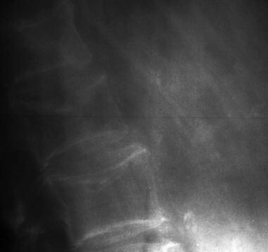