 |
VOOZH | about |
|
VOOZH | about |
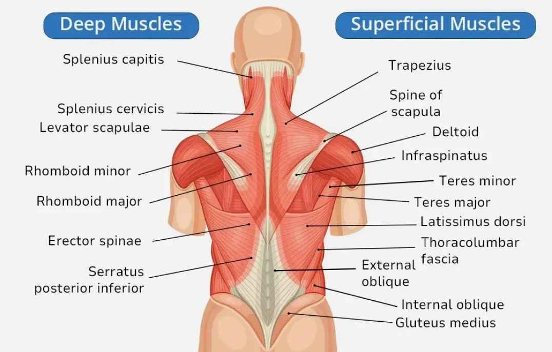
The back muscles are an intricate network that plays a vital role in maintaining posture, enabling movement, and safeguarding the spine. This dynamic system is made up of bones, tendons, ligaments, and muscles, each working together to support daily activities and protect the body’s core. The arrangement and interconnectivity of these muscles are essential for both stability and mobility.
The back muscles diagram illustrates the following structure:
Muscle Fibers
Muscle Layers
Attachment Points
Tendon Structure
Innervation
Movement
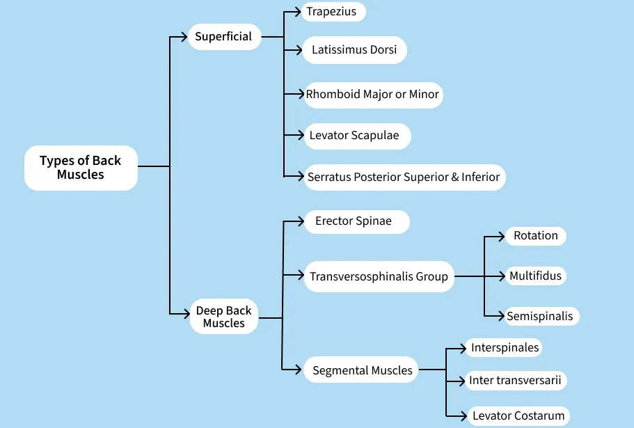
Back Muscles are divided into two parts, which are shown below:
The superficial back muscles are located closest to the surface of the body, making them easily accessible and often the most visible. These muscles are primarily involved in movements of the shoulder, arm, and upper trunk, as well as contributing to the overall stability and posture of the body.
The trapezius is one of the largest and most prominent muscles in the back. It spans the upper back, from the base of the skull to the middle of the spine and out to the shoulder blades. The trapezius plays a key role in the movement and stabilisation of the shoulder girdle and upper spine.
Function:
Parts:
The trapezius is divided into three parts based on its function and location:
The latissimus dorsi, commonly known as the "lats," is a large, flat muscle that stretches across the lower back and wraps around the sides. It plays an essential role in the movement of the arms and shoulders.
Function:
Attachment Points:
The rhomboid muscles, located between the shoulder blades, are responsible for retracting the scapula (pulling the shoulder blades towards the spine). These muscles are located underneath the trapezius and are critical for proper posture and shoulder movement.
Function:
Rhomboid Major vs. Minor:
The levator scapulae is a small muscle that runs along the side of the neck, extending from the cervical spine to the upper portion of the scapula. It is primarily involved in lifting the scapula.
Function:
These two smaller muscles are located at the top and bottom of the back and are involved in respiratory functions. They are relatively thin, and their actions are less obvious than the larger back muscles, but they still play a crucial role in movement and stabilisation.
Serratus Posterior Superior:
Serratus Posterior Inferior:
The deep back muscles are located beneath the superficial muscles and are primarily responsible for stabilising and supporting the spine. These muscles play an essential role in maintaining posture, enabling fine motor control, and facilitating complex movements of the spine. The deep back muscles are categorised into several groups based on their location and function, which include:
The erector spinae is a large muscle group that runs along the length of the spine, from the lower back to the neck. It is the most prominent group of deep back muscles and is primarily responsible for the extension and lateral flexion of the spine.
Structure: The erector spinae is divided into three main columns (or "groups") based on their location along the spine:
Function:
The transversospinalis group consists of smaller, deeper muscles that lie beneath the erector spinae. These muscles are responsible for stabilising and rotating the vertebral column. The main muscles in this group include the rotatores, multifidus, and semispinalis.
Transversospinalis group Muscles | Description | Functions |
|---|---|---|
1. Rotatores | These are the smallest muscles in the transversospinalis group and are located in the thoracic region. They span one or two vertebrae and are involved in the rotation and stabilisation of the spine. | Rotation and stabilisation of the vertebrae, especially in the thoracic and lumbar regions. |
2. Multifidus | The multifidus muscles are located throughout the entire vertebral column, from the sacrum to the cervical spine. They are the most significant deep spinal stabilisers, lying directly adjacent to the vertebrae. | They help in the stabilisation of the spine during movements, preventing excessive movement between vertebrae, and assisting in the rotation and extension of the spine. |
3. Semispinalis | The semispinalis muscles are located in the thoracic and cervical regions. They run from the transverse processes of the vertebrae to the spinous processes of the vertebrae higher up. | Primarily responsible for the extension and rotation of the spine, particularly in the cervical and upper thoracic regions. |
The segmental muscles are the smallest deep muscles of the back. They include the interspinales, intertransversarii, and levator costarum muscles. These muscles are responsible for fine-tuning the movements of the spine and providing additional stability.
Types of Segmental Muscles | Description | Function |
|---|---|---|
1. Interspinales | Located between adjacent spinous processes, these muscles assist in stabilising and extending the spine. | Aid in spinal extension and fine adjustments to the posture. |
2. Intertransversarii | These muscles lie between the transverse processes of adjacent vertebrae. They are found in both the cervical and lumbar regions. | Help in the lateral flexion of the spine and stabilise it during movement. |
3. Levator Costarum | These muscles originate from the transverse processes of the thoracic vertebrae and attach to the ribs. | Assist in elevating the ribs during respiration and contribute to the lateral flexion of the spine. |
The deep back muscles are crucial for both movement and stability of the spine. Their primary functions include:
Back muscles, like all muscles in the body, are made up of several key components that work together to generate force and movement. Here's a breakdown of the main components that make up the back muscles:
Back muscles are primarily composed of muscle fibres (also called muscle cells or myocytes). These fibres are elongated and cylindrical, capable of contracting and generating force when stimulated by nerve impulses. Muscle fibres are categorised into two main types:
Muscle fibres are grouped into bundles known as fascicles. A fascicle is a collection of muscle fibres surrounded by a layer of connective tissue called the perimysium. This organisation allows the muscles to work together efficiently while providing structural integrity.
The back muscles are surrounded and supported by various types of connective tissue, which help bind the fibres and fascicles together:
The back muscles are connected to bones through tendons. Tendons are tough, fibrous bands of connective tissue that transmit the force generated by the muscles to the bones, enabling movement. Tendons in the back can be either:
Back muscles are innervated by spinal nerves, which branch out from the spinal cord. These nerves carry electrical impulses that stimulate the muscle fibres to contract. The coordination of muscle contractions allows for controlled movement and stability. The nerves also provide sensory feedback to the brain, enabling proprioception (awareness of body position).
Like all muscles, the back muscles are supplied with oxygen and nutrients via blood vessels. The blood vessels provide essential oxygen and nutrients that fuel muscle contractions and aid in the removal of metabolic waste products (like carbon dioxide and lactic acid). The vascular system also helps regulate muscle temperature and maintain muscle function.
At a microscopic level, the muscle fibres are made up of smaller structures called myofibrils, which are long, thread-like structures that contain the actual contractile units of the muscle. Myofibrils are divided into repeated sections known as sarcomeres, which are the basic functional units of muscle contraction. The sarcomere contains two main protein filaments:
The interaction between actin and myosin within the sarcomeres leads to muscle contraction, which is the basis for the muscle's ability to move.
Back muscles serve several vital functions in the body, including:
Different types of muscle and back injuries occur due to strain, poor posture, ageing, or sudden movements. The following table shows common muscle injuries along with their causes and symptoms.
Muscle injuries | Causes | Symptoms |
|---|---|---|
1. Muscle Strain | Overstretching, lifting improperly, or sudden movements. | Sharp pain, muscle stiffness, swelling, and spasms. |
2. Sprain | Overstretched or torn ligaments, often from twisting or lifting. | Pain, swelling, limited movement, and instability. |
3. Herniated Disc | Disc bulging or rupturing, often due to ageing or injury. | Radiating pain, numbness, tingling, and muscle weakness. |
4. Sciatica | Pressure on the sciatic nerve, often from a herniated disc. | Pain radiating from the lower back down the leg, numbness, and weakness. |
5. Muscle Spasms | Overuse, dehydration, poor posture, or stress. | Suddenly, sharp pain and tightness in the back. |
6. Sacroiliac Joint Dysfunction | Injury, pregnancy, or poor posture. | Pain in the lower back or buttocks, worsened by movement. |
7. Facet Joint Dysfunction | Degeneration or trauma to the spine. | Localised pain, stiffness, and pain radiating to the hips or thighs. |
8. Spinal Stenosis | Narrowing of the spinal canal, usually from ageing. | Back or leg pain, numbness, and difficulty walking. |