 |
VOOZH | about |
|
VOOZH | about |
NCERT Notes of Class 12 Chapter 2 Human Reproduction: Human reproduction is the biological process by which a new individual offspring is produced from one or two parent organisms. The Human Reproduction process involves the fusion of gametes, which are specialized cells that carry genetic information from each parent, resulting in the formation of a zygote.
Human reproduction encompasses the whole process from fertilization to childbirth. Humans are sexually reproducing, internally fertilizing, and viviparous organisms. The reproductive system of humans includes the primary sex organs that produce the gametes, and secondary organs that show secondary sexual characteristics that separate males from females and help in the whole process of reproduction. We humans are sexually dimorphic i.e. there is a remarkable difference between male and female reproductive events, and these events only occur when a person achieves puberty. The events include the following stages;
The human Reproductive system is divided into two types i.e., Male and Female Reproductive Systems:
The male reproductive system is mostly located externally in the pelvic region of the body. It consists of the testes (primary sex organ), accessory ducts, accessory glands, and external genitalia.
It is the external pouch in which the testis is situated outside the body to maintain a temperature of 2 to 2.5°C lower than the body’s normal temperatures as the body’s temperature is not suitable for the formation of sperm cells. The scrotum is connected to the abdominal cavity and the pelvis with the help of the inguinal canal through which the spermatic cord passes.
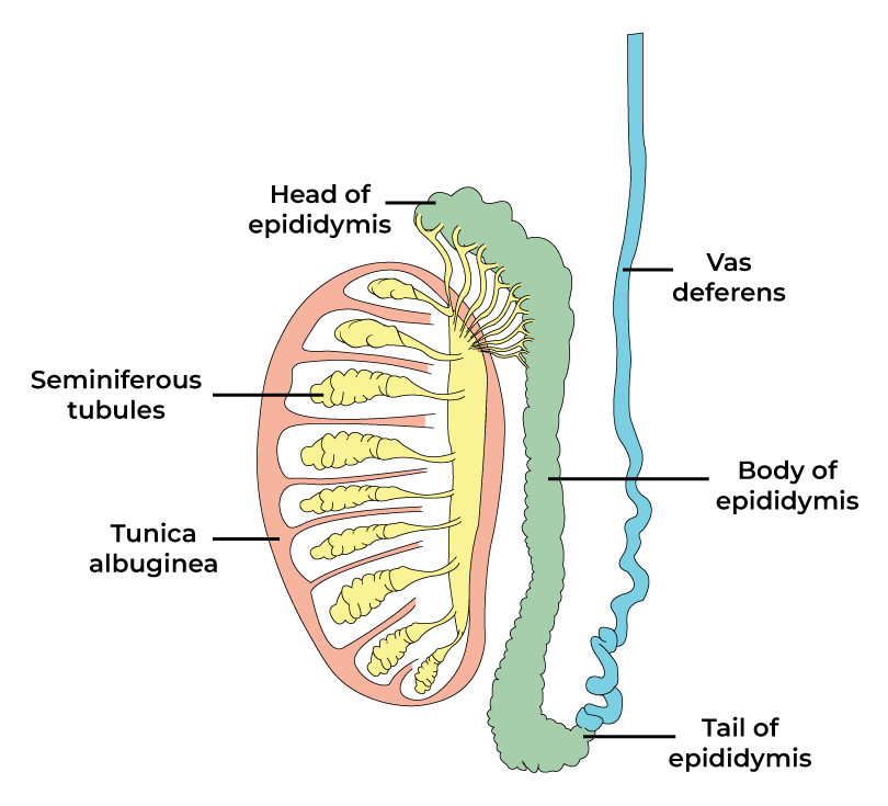
The male testis is the primary sex organ in the male reproductive system that is also called the male gonad. It is oval, 4-5 cm in length, 2-3 cm in width, and is covered by a dense covering (made of three layers i.e. tunica vaginalis, tunica albuginea, and tunica vasculosa).
Inside each testis, there are 250 compartments found which are called testicular lobules and each lobule consists of about 1 to 3 highly coiled tubular structures called the seminiferous tubules (site of sperm cell synthesis). The seminiferous tubules inside are lined by;
Outside the seminiferous tubules, there is the interstitial space present that consists of the blood vessels, and Leidig cells or the interstitial cells that are responsible for the secretion of male androgen i.e. testosterone.
The male duct system includes; rete testis, vasa efferentia, epididymis, and vas deferens. The pathway includes;
Seminiferous tubules (inside the testis) --> Rete testis (inside the testis) --> Vasa efferentia (inside the testis) --> Epididymis (present in the posterior surface of testis) --> Vas deferens (leads to the abdomen) --> Seminal duct (from seminal vesicle) + vas deferens + urethra --> Forms the ejaculatory duct.
The vas deferens form a loop over the urinary bladder. This duct system is responsible for the transport of sperm cells outside through an external opening called the urethral meatus.
It includes a seminal vesicle, a prostate, and Cowper’s glands.
It is commonly called the Penis which serves as both the reproductive organ as well as the urinal duct. There are three cylindrical masses of erectile tissue (two corpora cavernosa on the dorsal side, and one corpus spongiosum on the ventral side) that is responsible for the erection of the penis during copulation. The urethra passes through the corpus spongiosum and ends in the enlarged glans penis which is covered by the foreskin or prepuce.
It is the collection of secretions from the seminal vesicle, prostate, Cowper’s gland, and sperm cells. Its pH is around 7.3 to 7.5 i.e. slightly alkaline. In one ejaculate it consists of around 200 to 300 million sperm cells.
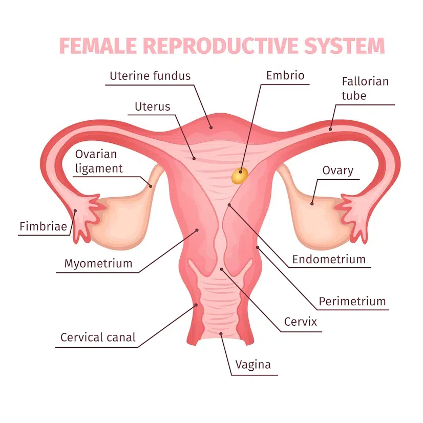
Female Reproductive System is located internally in the pelvic region of the lower abdomen. It consists of a pair of ovaries (primary sex organ), a pair of oviducts, a uterus, a cervix, a vagina, and external genitalia. Along with these structures, there is a pair of mammary glands that acts in a pattern to maintain the process of ovulation, fertilization, pregnancy, parturition, and lactation.
Ovaries are a pair of almond-shaped female gonads that is 2 to 4 cm in length, 1.5 cm in width, and 1 cm in thickness. It is attached to the uterine wall with the help of ovarian ligaments and is covered by a layer of cuboidal epithelium called the germinal epithelium which forms the oogonia. Below the epithelium is a layer of connective tissue called the tunica albuginea which covers the ovarian stroma. The stroma is divided into two regions- the outer cortex and the inner medulla.
It comprises a pair of fallopian tubes or oviducts, a uterus, and a vagina. Each oviduct is a 10 to 12-cm long, hollow tubular ciliated structure that extends from the periphery of the ovary to the uterus and is divided into three regions;
The uterus or the womb is a single, inverted pear-like structure that is attached to the pelvic wall with the help of ligaments. Its wall is made up of three layers of tissue;
The uterus opens through a narrow cervix into the vagina. The cervical canal of the cervix along with the vagina is called the birth canal. The vagina is about 10 cm long that extends from the cervix to the external genitalia. It is responsible for acting as the birth canal, allowing the menstrual flow, and accepting the male penis during copulation. The opening of the vagina is called the vaginal orifice which is covered partially by a membrane called the hymen.
There are mainly two categories of glands in the female reproductive system; the vestibular gland and the mammary gland. The vestibular gland is of two types;
The mammary glands or breasts are a pair of glandular tissue-containing structures that have a variable amount of fat in them. The glandular tissue of each breast is divided into 15 to 20 mammary lobes that contain a cluster of milk-secreting cells called alveoli. The milk is then stored in the cavities of alveoli which open into the mammary tubules that join to form the mammary duct. And several mammary ducts join to form the mammary ampulla that is connected to the lactiferous duct through which the milk comes out.
The process of formation of gametes (sperm and ovum) by meiosis in the primary sex organs i.e. testis in males and ovaries in females is called gametogenesis. The formation of sperm by the testis is called spermatogenesis and the formation of the ovum by ovaries is called oogenesis.
The meiotic process by which the four haploid spermatids having 23 chromosomes are produced from the diploid male germ cells having 46 chromosomes inside the seminiferous tubules of the testis is called spermatogenesis. It begins at the time of puberty. It has the following phases;
The spermatids will then undergo spermiogenesis in which the transformation of spermatids into spermatozoa (sperms) takes place. After spermiogenesis, sperm heads get embedded in the Sertoli cells and are finally released from the seminiferous tubules by the process called spermiation. The human male ejaculates about 200 to 300 million sperms during coitus in which, for normal fertility, at least 60 % of sperm cells must have normal shape and size and at least 40 % of them must show vigorous motility.
A mature sperm is a 0.06mm long, microscopic structure whose whole body is enveloped by a layer of the plasma membrane. Its body is composed of a head, neck, middle piece, and tail.
When a male reaches puberty the process of spermatogenesis starts due to the significant increase in the secretion of the Gonadotropin-releasing hormone (GnRH), which then acts on the anterior pituitary gland and stimulates the secretion of two gonadotropins; luteinizing hormone (LH) and follicle-stimulating hormone (FSH).
The process by which a mature female gamete or ovum is formed is called Oogenesis. Unlike making gametogenesis this process gets initiated during the embryonic development stage and includes the following phases;
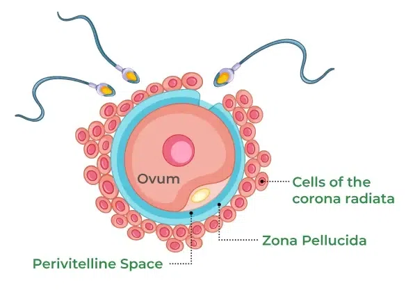
The ovum is a round, non-motile, and haploid cell that has a centrally located nucleus and dense cytoplasm. The cytoplasm stores food material required for the entire process of development. The ovum has four layers of covering; the plasma membrane, the Vitelline membrane, the zona pellucida, and the corona radiata.
The hypothalamus secretes the GnRH to stimulate the pituitary gland for the secretion of luteinizing hormone and follicle-stimulating hormone. LH stimulates the corpus luteum to secrete progesterone which when rises to undesirable levels inhibits the release of GnRH thus stopping the whole process. FSH stimulates the formation of estrogen and the development of the primary oocyte to form a secondary oocyte.
It is the reproductive cycle in female primates e.g. monkeys, apes, and human beings. The first menstruation begins at puberty and is called menarche. Menstrual cycles cease around 50 years of age; that is termed menopause. In human females, menstruation is repeated at an average interval of about 28/29 days, and the cycle of events starting from one menstruation to the next one is called the menstrual cycle. It has four phases;
During copulation (coitus) insemination takes place i.e. semen containing sperm cells is released by the penis into the vagina. The motile sperms swim rapidly, pass through the cervix, enter into the uterus, and finally reach the junction of the isthmus and ampulla (ampullary-isthmic junction) of the fallopian tube. On the other hand, the ovum released by the ovary is also transported to the ampullary-isthmic junction where fertilization takes place. For the process of fertilization to take place the sperm cells first have to undergo capacitation in which the sperm cells get activated by the secretion of the female genital tract for the release of enzymes stored in the acrosome.
Now fertilization i.e. fusion of male and female gamete will take place. It occurs only when the ovum and sperms are transported simultaneously to the ampullary-isthmic junction failing to do will miss fertilization and pregnancy. During fertilization, a sperm comes in contact with the zona pellucida layer of the ovum, secrets enzymes from the acrosome for its entry while inducing changes in the membrane that block the entry of additional sperms (Prevent polyspermy) thus ensuring only one sperm cell fertilizes an ovum. This induces the completion of the meiotic division of the secondary oocyte. The second meiotic division is also unequal and results in the formation of a small second polar body and a haploid egg. Soon the haploid nucleus of the sperm and that of the ovum will fuse to form a diploid zygote.
The mitotic division called the cleavage will start as the zygote moves towards the uterus through the isthmus and forms 2, 4, 8, 16 celled daughter cells called blastomeres. The embryo with 8 to 16 blastomeres is called a morula which will continue to divide and transforms into a blastocyst. In the blastocyst, the blastomeres are arranged into an outer layer called trophoblast (later get attached as finger-like projections called chorionic villi to the endometrium) and an inner group of cells called inner cell mass (give rise to the embryo ) attached to trophoblast. After attachment, the uterine cell divides rapidly and covers the blastocyst embedding it in the endometrium of the uterus. This is called implantation and it leads to pregnancy.
Fusion of the male and female nuclei will determine the sex of the baby. As the chromosomal pattern in human females is XX and males is XY, the gametes will be X in the case of females whereas male gametes will either be X or Y. When the sperm cell carrying an X chromosome will fertilize the female ovum the zygote will have XX i.e. it will be a female child and when the Y chromosome carrying sperm will fertilize the ovum the zygote will be XY i.e. the baby will be a male.
Immediately after implantation, the inner cell mass (embryo) differentiates into an outer layer called ectoderm and an inner layer called endoderm, and in between a mesoderm soon appears. These three layers give rise to all tissues (organs) in adults.
After implantation, the chorionic villi (finger-like projections) get surrounded by the uterine tissue and maternal blood. The chorionic villi and uterine tissue become interdigitated with each other and jointly form a structural and functional unit between the developing embryo (fetus) and maternal body called the placenta that is connected to the embryo through an umbilical cord and helps in the transport of substances to and from the embryo. The functions of the placenta are;
The duration between fertilization and parturition (9 months) is called gestation. During gestation following stages of development are seen;
Vigorous contraction of the uterus at the end of pregnancy causes expulsion/delivery of the fetus is called parturition. It is controlled by a neuroendocrine mechanism that includes the following steps;
Soon after the infant is delivered, the placenta is also expelled out of the uterus. The production of milk from the mammary glands is called lactation. The mammary glands of the female undergo differentiation during pregnancy and start producing milk towards the end of pregnancy. This helps the mother in feeding the newborn. The milk produced during the initial few days of lactation is called the colostrum which contains several antibodies essential to develop resistance or immunity in newborn babies.