
- FASTA Sequence
- PDBx/mmCIF Format
- PDBx/mmCIF Format (gz)
- BinaryCIF Format (gz)
- Legacy PDB Format
- Legacy PDB Format (gz)
- PDBML/XML Format (gz)
- NMR Restraints (Text)
- NMR Restraints (Text - gz)
- NMR Restraints v2 (Text)
- NMR Restraints v2 (Text - gz)
- Validation Full (PDF - gz)
- Validation (XML - gz)
- Validation (CIF - gz)
- Biological Assembly 1 (CIF - gz)
- Biological Assembly 1 (PDB - gz)
1B22 | pdb_00001b22
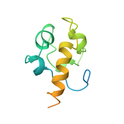
RAD51 (N-TERMINAL DOMAIN)
- PDB DOI: https://doi.org/10.2210/pdb1B22/pdb
- BMRB: 4328
- Classification: DNA BINDING PROTEIN
- Organism(s): Homo sapiens
- Expression System: Escherichia coli
- Mutation(s): No
- Deposited: 1998-12-04 Released: 1999-12-03
- Deposition Author(s): Aihara, H., Ito, Y., Kurumizaka, H., Yokoyama, S., Shibata, T., RIKEN Structural Genomics/Proteomics Initiative (RSGI)
Experimental Data Snapshot
- Method: SOLUTION NMR
- Conformers Calculated: 140
- Conformers Submitted: 30
- Selection Criteria: LEAST TARGET FUNCTION VALUES
wwPDB Validation 3D Report Full Report
- 👁 Image
Download Mendeley
The N-terminal domain of the human Rad51 protein binds DNA: structure and a DNA binding surface as revealed by NMR.
Aihara, H., Ito, Y., Kurumizaka, H., Yokoyama, S., Shibata, T.(1999) J Mol Biology 290: 495-504
- PubMed: 10390347 Search on PubMed
- DOI: https://doi.org/10.1006/jmbi.1999.2904
- Primary Citation Related Structures:
1B22 - PubMed Abstract:
Human Rad51 protein (HsRad51) is a homolog of Escherichia coli RecA protein, and functions in DNA repair and recombination. In higher eukaryotes, Rad51 protein is essential for cell viability. The N-terminal region of HsRad51 is highly conserved among eukaryotic Rad51 proteins but is absent from RecA, suggesting a Rad51-specific function for this region. Here, we have determined the structure of the N-terminal part of HsRad51 by NMR spectroscopy. The N-terminal region forms a compact domain consisting of five short helices, which shares structural similarity with a domain of endonuclease III, a DNA repair enzyme of E. coli. NMR experiments did not support the involvement of the N-terminal domain in HsRad51-HsBrca2 interaction or the self-association of HsRad51 as proposed by previous studies. However, NMR tiration experiments demonstrated a physical interaction of the domain with DNA, and allowed mapping of the DNA binding surface. Mutation analysis showed that the DNA binding surface is essential for double-stranded and single-stranded DNA binding of HsRad51. Our results suggest the presence of a DNA binding site on the outside surface of the HsRad51 filament and provide a possible explanation for the regulation of DNA binding by phosphorylation within the N-terminal domain.
- Cellular & Molecular Biology Laboratory, The Institute of Physical and Chemical Research (RIKEN), 2-1 Hirosawa, Wako-shi, Saitama, 351-0198, Japan.
Organizational Affiliation:
Explore in 3D: Structure | Sequence Annotations | Validation Report
Global Symmetry: Asymmetric - C1
Global Stoichiometry: Monomer - A1
Find Similar Assemblies
Biological assembly 1 assigned by authors.
Macromolecule Content
- Total Structure Weight: 12.55 kDa
- Atom Count: 528
- Modeled Residue Count: 70
- Deposited Residue Count: 114
- Unique protein chains: 1
Entity ID: 1 | |||||
|---|---|---|---|---|---|
| Molecule | Chains | Sequence Length | Organism | Details | Image |
| DNA REPAIR PROTEIN RAD51 | 114 | Homo sapiens | Mutation(s): 0 EC: 3.6.4 | 👁 Image | |
UniProt & NIH Common Fund Data Resources | |||||
Find proteins for Q06609 (Homo sapiens) Explore Q06609 Go to UniProtKB: Q06609 | |||||
PHAROS: Q06609 GTEx: ENSG00000051180 | |||||
Entity Groups | |||||
| Sequence Clusters | 30% Identity50% Identity70% Identity90% Identity95% Identity100% Identity | ||||
| UniProt Group | Q06609 | ||||
Sequence AnnotationsExpand | |||||
| |||||
Experimental Data
- Method: SOLUTION NMR
- Conformers Calculated: 140
- Conformers Submitted: 30
- Selection Criteria: LEAST TARGET FUNCTION VALUES
Deposition Data
- Released Date: 1999-12-03 Deposition Author(s): Aihara, H., Ito, Y., Kurumizaka, H., Yokoyama, S., Shibata, T., RIKEN Structural Genomics/Proteomics Initiative (RSGI)
Revision History (Full details and data files)
- Version 1.0: 1999-12-03
Type: Initial release - Version 1.1: 2008-04-26
Changes: Version format compliance - Version 1.2: 2011-07-13
Changes: Version format compliance - Version 1.3: 2022-02-16
Changes: Database references, Derived calculations - Version 1.4: 2023-12-27
Changes: Data collection