
- FASTA Sequence
- PDBx/mmCIF Format
- PDBx/mmCIF Format (gz)
- BinaryCIF Format (gz)
- Legacy PDB Format
- Legacy PDB Format (gz)
- PDBML/XML Format (gz)
- Validation Full (PDF - gz)
- Validation (XML - gz)
- Validation (CIF - gz)
- Biological Assembly 1 (CIF - gz)
- Biological Assembly 2 (CIF - gz)
- Biological Assembly 1 (PDB - gz)
- Biological Assembly 2 (PDB - gz)
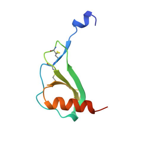
1DOK | pdb_00001dok
MONOCYTE CHEMOATTRACTANT PROTEIN 1, P-FORM
- PDB DOI: https://doi.org/10.2210/pdb1DOK/pdb
- Classification: CHEMOATTRACTANT
- Organism(s): Homo sapiens
- Expression System: Escherichia coli
- Mutation(s): No
- Deposited: 1996-11-27 Released: 1997-03-12
- Deposition Author(s): Lubkowski, J., Bujacz, G., Boque, L., Wlodawer, A., Domaille, P.J., Handel, T.M.
Experimental Data Snapshot
- Method: X-RAY DIFFRACTION
- Resolution: 1.85 Å
- R-Value Free: 0.245 (Depositor)
- R-Value Work: 0.191 (Depositor)
- R-Value Observed: 0.191 (Depositor)
Starting Model: experimental
View more details
wwPDB Validation3D Report Full Report
Literature
- 👁 Image
Download Mendeley
The structure of MCP-1 in two crystal forms provides a rare example of variable quaternary interactions.
Lubkowski, J., Bujacz, G., Boque, L., Domaille, P.J., Handel, T.M., Wlodawer, A.(1997) Nat Struct Biol 4: 64-69
- PubMed: 8989326 Search on PubMed
- DOI: https://doi.org/10.1038/nsb0197-64
- Primary Citation Related Structures:
1DOK, 1DOL - PubMed Abstract:
The X-ray crystal structure of recombinant human monocyte chemoattractant protein (MCP-1) has been solved in two crystal forms. One crystal form (P), refined to 1.85 A resolution, contains a dimer in the asymmetric unit, while the other (I) contains a monomer and was refined at 2.4 A. Although both crystal forms grow together in the same droplet, the respective quaternary structures of the protein differ dramatically. In addition, both X-ray structures differ to a similar extent from the solution structure of MCP-1. Such extent of variability of quaternary structures is unprecedented. In the crystal structures, the well-ordered N termini of MCP-1 form 3(10)-helices. Comparison of the three MCP-1 structures revealed a direct correlation between the main-chain conformation of the first two cysteine residues and the quaternary arrangements. These data can be used to explain the structural basis for the assignment of residues responsible for biological activity.
- Macromolecular Structure Laboratory, NCI-Frederick Cancer Research and Development Center, ABL-Basic Research Program, Maryland 21702, USA.
Organizational Affiliation:
Explore in 3D: Structure | Sequence Annotations | Validation Report | Ligand Interaction (SO4)
Biological Assembly 1
Explore in 3D: Structure | Sequence Annotations | Validation Report | Ligand Interaction (SO4)
Global Symmetry: Cyclic - C2 (Explore in 3D)
Global Stoichiometry: Homo 2-mer - A2
Find Similar Assemblies
Biological assembly 1 assigned by authors and generated by PISA (software)
Biological Assembly 2
Explore in 3D: Structure | Sequence Annotations | Validation Report | Ligand Interaction (SO4)
Global Symmetry: Cyclic - C2 (Explore in 3D)
Global Stoichiometry: Homo 4-mer - A4
Find Similar Assemblies
Biological assembly 2 generated by PISA (software)
Macromolecule Content
- Total Structure Weight: 17.85 kDa
- Atom Count: 1,325
- Modeled Residue Count: 144
- Deposited Residue Count: 154
- Unique protein chains: 1
Macromolecules
Entity ID: 1 | |||||
|---|---|---|---|---|---|
| Molecule | Chains | Sequence Length | Organism | Details | Image |
| MONOCYTE CHEMOATTRACTANT PROTEIN 1 | 77 | Homo sapiens | Mutation(s): 0 | 👁 Image | |
UniProt & NIH Common Fund Data Resources | |||||
PHAROS: P13500 GTEx: ENSG00000108691 | |||||
Entity Groups | |||||
| Sequence Clusters | 30% Identity50% Identity70% Identity90% Identity95% Identity100% Identity | ||||
| UniProt Group | P13500 | ||||
Sequence AnnotationsExpand | |||||
Reference Sequence | |||||
Small Molecules
| Ligands 1 Unique | |||||
|---|---|---|---|---|---|
| ID | Chains | Name / Formula / InChI Key | 2D Diagram | 3D Interactions | |
| SO4 Download:Ideal Coordinates CCD File | C [auth A], D [auth B] | SULFATE ION O4 S QAOWNCQODCNURD-UHFFFAOYSA-L | 👁 Image | ||
Experimental Data & Validation
Experimental Data
- Method: X-RAY DIFFRACTION
- Resolution: 1.85 Å
- R-Value Free: 0.245 (Depositor)
- R-Value Work: 0.191 (Depositor)
- R-Value Observed: 0.191 (Depositor)
| Length ( Å ) | Angle ( ˚ ) |
|---|---|
| a = 50.74 | α = 90 |
| b = 50.74 | β = 90 |
| c = 107.65 | γ = 90 |
| Software Name | Purpose |
|---|---|
| DENZO | data reduction |
| SCALEPACK | data scaling |
| AMoRE | phasing |
| X-PLOR | refinement |
Entry History
Deposition Data
- Released Date: 1997-03-12 Deposition Author(s): Lubkowski, J., Bujacz, G., Boque, L., Wlodawer, A., Domaille, P.J., Handel, T.M.
Revision History (Full details and data files)
- Version 1.0: 1997-03-12
Type: Initial release - Version 1.1: 2008-03-24
Changes: Version format compliance - Version 1.2: 2011-07-13
Changes: Version format compliance - Version 1.3: 2023-08-09
Changes: Database references, Derived calculations, Refinement description - Version 1.4: 2024-10-16
Changes: Data collection, Structure summary