
- FASTA Sequence
- PDBx/mmCIF Format
- PDBx/mmCIF Format (gz)
- BinaryCIF Format (gz)
- Legacy PDB Format
- Legacy PDB Format (gz)
- PDBML/XML Format (gz)
- NMR Restraints (Text)
- NMR Restraints (Text - gz)
- NMR Restraints v2 (Text)
- NMR Restraints v2 (Text - gz)
- Validation Full (PDF - gz)
- Validation (XML - gz)
- Validation (CIF - gz)
- Biological Assembly 1 (CIF - gz)
- Biological Assembly 1 (PDB - gz)
1DW5 | pdb_00001dw5
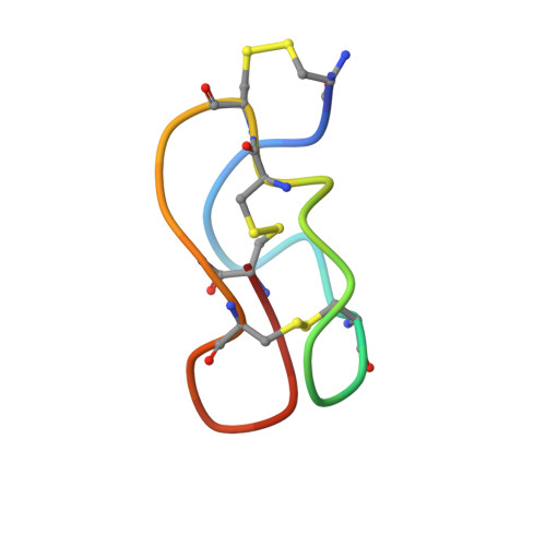
NMR STRUCTURE OF OMEGA-CONOTOXIN MVIIA: NO CONSTRAINTS ON DISULPHIDE BRIDGES
- PDB DOI: https://doi.org/10.2210/pdb1DW5/pdb
- Classification: TOXIN
- Organism(s): Conus magus
- Mutation(s): No
- Deposited: 2000-01-24 Released: 2000-03-01
- Deposition Author(s): Atkinson, R.A., Kieffer, B., Dejaegere, A., Sirockin, F., Lefevre, J.-F.
Experimental Data Snapshot
- Method: SOLUTION NMR
- Conformers Calculated: 500
- Conformers Submitted: 25
- Selection Criteria: structures with the lowest energy
wwPDB Validation 3D Report Full Report
- 👁 Image
Download Mendeley
Structural and dynamic characterization of omega-conotoxin MVIIA: the binding loop exhibits slow conformational exchange.
Atkinson, R.A., Kieffer, B., Dejaegere, A., Sirockin, F., Lefevre, J.F.(2000) Biochemistry 39: 3908-3919
- PubMed: 10747778 Search on PubMed
- DOI: https://doi.org/10.1021/bi992651h
- Primary Citation Related Structures:
1DW4, 1DW5 - PubMed Abstract:
omega-Conotoxin MVIIA is a 25-residue, disulfide-bridged polypeptide from the venom of the sea snail Conus magus that binds to neuronal N-type calcium channels. It forms a compact folded structure, presenting a loop between Cys8 and Cys15 that contains a set of residues critical for its binding. The loop does not have a unique defined structure, nor is it intrinsically flexible. Broadening of a subset of resonances in the NMR spectrum at low temperature, anomalous temperature dependence of the chemical shifts of some resonances, and exchange contributions to J(0) from (13)C relaxation measurements reveal that conformational exchange affects the residues in this loop. The effects of this exchange on the calculated structure of omega-conotoxin MVIIA are discussed. The exchange appears to be associated with a change in the conformation of the disulfide bridge Cys8-Cys20. The implications for the use of the omega-conotoxins as a scaffold for carrying other functions is discussed.
- UPR 9003 du CNRS, Ecole Supérieure de Biotechnologie de Strasbourg, Bld. Sébastien Brant, 67400 Illkirch, France. aatkins@nimr.mrc.ac.uk
Organizational Affiliation:
Explore in 3D: Structure | Sequence Annotations | Validation Report
Global Symmetry: Asymmetric - C1
Global Stoichiometry: Monomer - A1
Find Similar Assemblies
Biological assembly 1 assigned by authors.
Macromolecule Content
- Total Structure Weight: 2.65 kDa
- Atom Count: 177
- Modeled Residue Count: 26
- Deposited Residue Count: 26
- Unique protein chains: 1
Entity ID: 1 | |||||
|---|---|---|---|---|---|
| Molecule | Chains | Sequence Length | Organism | Details | Image |
| OMEGA-CONOTOXIN MVIIA | 26 | N/A | Mutation(s): 0 | 👁 Image | |
UniProt | |||||
Find proteins for P05484 (Conus magus) Explore P05484 Go to UniProtKB: P05484 | |||||
Entity Groups | |||||
| Sequence Clusters | 30% Identity50% Identity70% Identity90% Identity95% Identity100% Identity | ||||
| UniProt Group | P05484 | ||||
Sequence AnnotationsExpand | |||||
| |||||
Experimental Data
- Method: SOLUTION NMR
- Conformers Calculated: 500
- Conformers Submitted: 25
- Selection Criteria: structures with the lowest energy
Deposition Data
- Released Date: 2000-03-01 Deposition Author(s): Atkinson, R.A., Kieffer, B., Dejaegere, A., Sirockin, F., Lefevre, J.-F.
Revision History (Full details and data files)
- Version 1.0: 2000-03-01
Type: Initial release - Version 1.1: 2008-04-27
Changes: Version format compliance - Version 1.2: 2011-07-13
Changes: Version format compliance - Version 1.3: 2022-02-16
Changes: Data collection, Database references, Derived calculations - Version 1.4: 2024-11-20
Changes: Data collection, Structure summary