
- FASTA Sequence
- PDBx/mmCIF Format
- PDBx/mmCIF Format (gz)
- BinaryCIF Format (gz)
- Legacy PDB Format
- Legacy PDB Format (gz)
- PDBML/XML Format (gz)
- NMR Restraints (Text)
- NMR Restraints (Text - gz)
- NMR Restraints v2 (Text)
- NMR Restraints v2 (Text - gz)
- Validation Full (PDF - gz)
- Validation (XML - gz)
- Validation (CIF - gz)
- Biological Assembly 1 (CIF - gz)
- Biological Assembly 1 (PDB - gz)
1HRJ | pdb_00001hrj
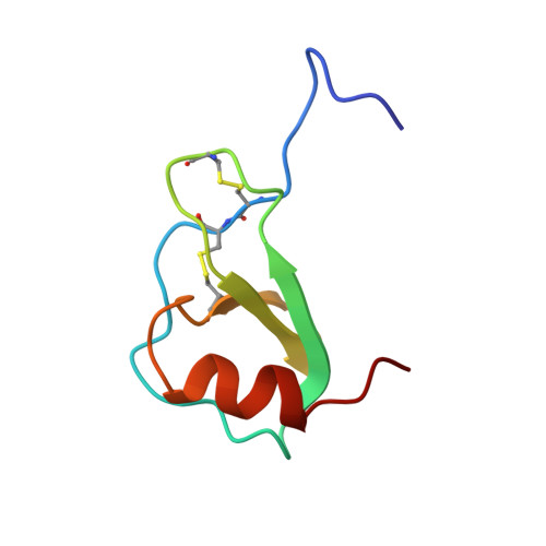
HUMAN RANTES, NMR, 13 STRUCTURES
- PDB DOI: https://doi.org/10.2210/pdb1HRJ/pdb
- Classification: CYTOKINE (CHEMOTACTIC)
- Organism(s): Homo sapiens
- Expression System: Escherichia coli
- Mutation(s): No
- Deposited: 1995-08-18 Released: 1996-10-14
- Deposition Author(s): Chung, C., Cooke, R.M., Proudfoot, A.E.I., Wells, T.N.C.
Experimental Data Snapshot
- Method: SOLUTION NMR
- Conformers Submitted: 13
wwPDB Validation3D Report Full Report
Literature
- 👁 Image
Download Mendeley
The three-dimensional solution structure of RANTES.
Chung, C.W., Cooke, R.M., Proudfoot, A.E., Wells, T.N.(1995) Biochemistry 34: 9307-9314
- PubMed: 7542919 Search on PubMed
- DOI: https://doi.org/10.1021/bi00029a005
- Primary Citation Related Structures:
1HRJ - PubMed Abstract:
The solution structure of the chemokine RANTES (regulated on activation, normal T-cell expressed and secreted) has been determined using NMR spectroscopy. Backbone and side-chain 1H and 15N assignments have been obtained using a combination of two-dimensional homonuclear and three-dimensional heteronuclear spectra. Regular elements of secondary structure have been identified on the basis of a qualitative interpretation of NOE data, J(NH-H alpha) coupling constants, and amide exchange rates. Three-dimensional structures were calculated from a total of 2146 experimental restraints using a combination of distance geometry and simulated annealing protocols. For the 13 best structures the average backbone (N, C alpha, C) atomic rmsd from the mean coordinates for residues 5-65 is 0.64 A (+/- 0.14 A) for the dimer and 0.50 A (+/- 0.08 A) for the individual monomers. Each monomer consists of a three-stranded antiparallel beta-sheet (residues 26-30, 38-43, 48-51) in a Greek key motif with a C-terminal helix (56-65) packed across the sheet, an arrangement similar to the monomeric structure of other members of this chemokine family (IL-8, PF4, MGSA/Gro alpha, and MIP-1 beta). Overall, the RANTES dimer resembles that previously reported for MIP-1 beta.
- Department of Biomolecular Structure, Glaxo Research and Development Ltd., Hertfordshire, U.K.
Organizational Affiliation:
Biological Assembly 1
Explore in 3D: Structure | Sequence Annotations | Validation Report
Global Symmetry: Cyclic - C2 (Explore in 3D)
Global Stoichiometry: Homo 2-mer - A2
Find Similar Assemblies
Biological assembly 1 assigned by authors.
Macromolecule Content
- Total Structure Weight: 15.72 kDa
- Atom Count: 1,102
- Modeled Residue Count: 136
- Deposited Residue Count: 136
- Unique protein chains: 1
Macromolecules
Entity ID: 1 | |||||
|---|---|---|---|---|---|
| Molecule | Chains | Sequence Length | Organism | Details | Image |
| HUMAN REGULATED UPON ACTIVATION NORMAL T-CELL EXPRESSED AND SECRETED | 68 | Homo sapiens | Mutation(s): 0 | 👁 Image | |
UniProt & NIH Common Fund Data Resources | |||||
PHAROS: P13501 GTEx: ENSG00000271503 | |||||
Entity Groups | |||||
| Sequence Clusters | 30% Identity50% Identity70% Identity90% Identity95% Identity100% Identity | ||||
| UniProt Group | P13501 | ||||
Sequence AnnotationsExpand | |||||
Reference Sequence | |||||
Experimental Data & Validation
Experimental Data
- Method: SOLUTION NMR
- Conformers Submitted: 13
Entry History
Deposition Data
- Released Date: 1996-10-14 Deposition Author(s): Chung, C., Cooke, R.M., Proudfoot, A.E.I., Wells, T.N.C.
Revision History (Full details and data files)
- Version 1.0: 1996-10-14
Type: Initial release - Version 1.1: 2008-03-24
Changes: Version format compliance - Version 1.2: 2011-07-13
Changes: Version format compliance - Version 1.3: 2022-02-23
Changes: Database references, Derived calculations, Other - Version 1.4: 2024-10-16
Changes: Data collection, Structure summary