
- FASTA Sequence
- PDBx/mmCIF Format
- PDBx/mmCIF Format (gz)
- BinaryCIF Format (gz)
- Legacy PDB Format
- Legacy PDB Format (gz)
- PDBML/XML Format (gz)
- Structure Factors (CIF)
- Structure Factors (CIF - gz)
- Validation Full (PDF - gz)
- Validation (XML - gz)
- Validation (CIF - gz)
- Validation 2fo-fc coefficients (CIF - gz)
- Validation fo-fc coefficients (CIF - gz)
- Biological Assembly 1 (CIF - gz)
- Biological Assembly 1 (PDB - gz)
1IAM | pdb_00001iam
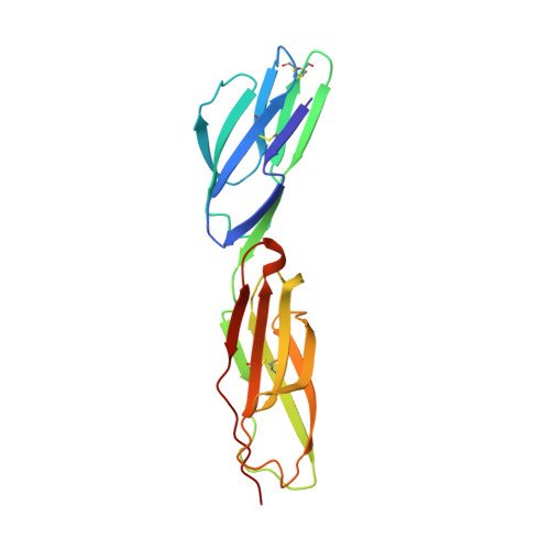
STRUCTURE OF THE TWO AMINO-TERMINAL DOMAINS OF HUMAN INTERCELLULAR ADHESION MOLECULE-1, ICAM-1
- PDB DOI: https://doi.org/10.2210/pdb1IAM/pdb
- Classification: Viral protein receptor
- Organism(s): Homo sapiens
- Expression System: Spodoptera frugiperda
- Mutation(s): Yes
- Deposited: 1998-02-22 Released: 1998-04-29
- Deposition Author(s): Bella, J., Kolatkar, P.R., Marlor, C., Greve, J.M., Rossmann, M.G.
Experimental Data Snapshot
- Method: X-RAY DIFFRACTION
- Resolution: 2.10 Å
- R-Value Free: 0.303 (Depositor)
- R-Value Work: 0.214 (Depositor), 0.230 (DCC)
- R-Value Observed: 0.214 (Depositor)
Starting Model: experimental
View more details
- 👁 Image
Download Mendeley
The structure of the two amino-terminal domains of human ICAM-1 suggests how it functions as a rhinovirus receptor and as an LFA-1 integrin ligand.
Bella, J., Kolatkar, P.R., Marlor, C.W., Greve, J.M., Rossmann, M.G.(1998) Proc Natl Acad Sci U S A 95: 4140-4145
- PubMed: 9539703 Search on PubMedSearch on PubMed Central
- DOI: https://doi.org/10.1073/pnas.95.8.4140
- Primary Citation Related Structures:
1IAM - PubMed Abstract:
The normal function of human intercellular adhesion molecule-1 (ICAM-1) is to provide adhesion between endothelial cells and leukocytes after injury or stress. ICAM-1 binds to leukocyte function-associated antigen (LFA-1) or macrophage-1 antigen (Mac-1). However, ICAM-1 is also used as a receptor by the major group of human rhinoviruses and is a catalyst for the subsequent viral uncoating during cell entry. The three-dimensional atomic structure of the two amino-terminal domains (D1 and D2) of ICAM-1 has been determined to 2.2-A resolution and fitted into a cryoelectron microscopy reconstruction of a rhinovirus-ICAM-1 complex. Rhinovirus attachment is confined to the BC, CD, DE, and FG loops of the amino-terminal Ig-like domain (D1) at the end distal to the cellular membrane. The loops are considerably different in structure to those of human ICAM-2 or murine ICAM-1, which do not bind rhinoviruses. There are extensive charge interactions between ICAM-1 and human rhinoviruses, which are mostly conserved in both major and minor receptor groups of rhinoviruses. The interaction of ICAMs with LFA-1 is known to be mediated by a divalent cation bound to the insertion (I)-domain on the alpha chain of LFA-1 and the carboxyl group of a conserved glutamic acid residue on ICAMs. Domain D1 has been docked with the known structure of the I-domain. The resultant model is consistent with mutational data and provides a structural framework for the adhesion between these molecules.
- Department of Biological Sciences, Purdue University, West Lafayette, IN 47907-1392, USA.
Organizational Affiliation:
Explore in 3D: Structure | Sequence Annotations | Electron Density | Validation Report | Ligand Interaction (NAG)
Explore in 3D: Structure | Sequence Annotations | Electron Density | Validation Report | Ligand Interaction (NAG)
Global Symmetry: Cyclic - C2 (Explore in 3D)
Global Stoichiometry: Homo 2-mer - A2
Find Similar Assemblies
Biological assembly 1 assigned by authors.
Macromolecule Content
- Total Structure Weight: 20.7 kDa
- Atom Count: 1,597
- Modeled Residue Count: 185
- Deposited Residue Count: 185
- Unique protein chains: 1
Entity ID: 1 | |||||
|---|---|---|---|---|---|
| Molecule | Chains | Sequence Length | Organism | Details | Image |
| INTERCELLULAR ADHESION MOLECULE-1 | 185 | Homo sapiens | Mutation(s): 3 | 👁 Image | |
UniProt & NIH Common Fund Data Resources | |||||
Find proteins for P05362 (Homo sapiens) Explore P05362 Go to UniProtKB: P05362 | |||||
PHAROS: P05362 GTEx: ENSG00000090339 | |||||
Entity Groups | |||||
| Sequence Clusters | 30% Identity50% Identity70% Identity90% Identity95% Identity100% Identity | ||||
| UniProt Group | P05362 | ||||
Glycosylation | |||||
| Glycosylation Sites: 1 | Go to GlyGen: P05362-1 | ||||
Sequence AnnotationsExpand | |||||
| |||||
| Ligands 1 Unique | |||||
|---|---|---|---|---|---|
| ID | Chains | Name / Formula / InChI Key | 2D Diagram | 3D Interactions | |
| NAG Query on NAG Download Ideal Coordinates CCD File | B [auth A] | 2-acetamido-2-deoxy-beta-D-glucopyranose C8 H15 N O6 OVRNDRQMDRJTHS-FMDGEEDCSA-N | 👁 Image | ||
Experimental Data
- Method: X-RAY DIFFRACTION
- Resolution: 2.10 Å
- R-Value Free: 0.303 (Depositor)
- R-Value Work: 0.214 (Depositor), 0.230 (DCC)
- R-Value Observed: 0.214 (Depositor)
| Length ( Å ) | Angle ( ˚ ) |
|---|---|
| a = 41.857 | α = 90 |
| b = 124.736 | β = 90 |
| c = 83.186 | γ = 90 |
| Software Name | Purpose |
|---|---|
| DENZO | data reduction |
| SCALEPACK | data scaling |
| X-PLOR | model building |
| X-PLOR | refinement |
| X-PLOR | phasing |
Deposition Data
- Released Date: 1998-04-29 Deposition Author(s): Bella, J., Kolatkar, P.R., Marlor, C., Greve, J.M., Rossmann, M.G.
Revision History (Full details and data files)
- Version 1.0: 1998-04-29
Type: Initial release - Version 1.1: 2008-03-24
Changes: Version format compliance - Version 1.2: 2011-07-13
Changes: Non-polymer description, Version format compliance - Version 1.3: 2011-11-16
Changes: Atomic model - Version 1.4: 2020-07-29
Type: Remediation
Reason: Carbohydrate remediation
Changes: Data collection, Derived calculations, Structure summary - Version 1.5: 2021-11-03
Changes: Database references, Structure summary - Version 1.6: 2023-08-09
Changes: Refinement description - Version 1.7: 2024-10-30
Changes: Data collection, Structure summary