
- FASTA Sequence
- PDBx/mmCIF Format
- PDBx/mmCIF Format (gz)
- BinaryCIF Format (gz)
- Legacy PDB Format
- Legacy PDB Format (gz)
- PDBML/XML Format (gz)
- Structure Factors (CIF)
- Structure Factors (CIF - gz)
- Validation Full (PDF - gz)
- Validation (XML - gz)
- Validation (CIF - gz)
- Validation 2fo-fc coefficients (CIF - gz)
- Validation fo-fc coefficients (CIF - gz)
- Biological Assembly 1 (CIF - gz)
- Biological Assembly 2 (CIF - gz)
- Biological Assembly 1 (PDB - gz)
- Biological Assembly 2 (PDB - gz)
1IC1 | pdb_00001ic1
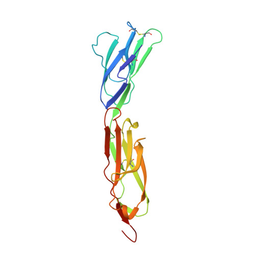
THE CRYSTAL STRUCTURE FOR THE N-TERMINAL TWO DOMAINS OF ICAM-1
- PDB DOI: https://doi.org/10.2210/pdb1IC1/pdb
- Classification: CELL ADHESION
- Organism(s): Homo sapiens
- Expression System: Cricetulus griseus
- Mutation(s): No
- Deposited: 1998-03-09 Released: 1998-06-17
- Deposition Author(s): Casasnovas, J.M., Stehle, T., Liu, J.-H., Wang, J.-H., Springer, T.A.
Experimental Data Snapshot
- Method: X-RAY DIFFRACTION
- Resolution: 3.00 Å
- R-Value Free: 0.279 (Depositor)
- R-Value Work: 0.222 (Depositor), 0.240 (DCC)
- R-Value Observed: 0.222 (Depositor)
Starting Models: experimental
View more details
- 👁 Image
Download Mendeley
A dimeric crystal structure for the N-terminal two domains of intercellular adhesion molecule-1.
Casasnovas, J.M., Stehle, T., Liu, J.H., Wang, J.H., Springer, T.A.(1998) Proc Natl Acad Sci U S A 95: 4134-4139
- PubMed: 9539702 Search on PubMedSearch on PubMed Central
- DOI: https://doi.org/10.1073/pnas.95.8.4134
- Primary Citation Related Structures:
1IC1 - PubMed Abstract:
The 3.0-A structure of a 190-residue fragment of intercellular adhesion molecule-1 (ICAM-1, CD54) reveals two tandem Ig-superfamily (IgSF) domains. Each of two independent molecules dimerizes identically with a symmetry-related molecule over a hydrophobic interface on the BED sheet of domain 1, in agreement with dimerization of ICAM-1 on the cell surface. The residues that bind to the integrin LFA-1 are well oriented for bivalent binding in the dimer, with the critical Glu-34 residues pointing away from each other on the periphery. Residues that bind to rhinovirus are in the flexible BC and FG loops at the tip of domain 1, and these and the upper half of domain 1 are well exposed in the dimer for docking to virus. By contrast, a residue important for binding to Plasmodium falciparum-infected erythrocytes is in the dimer interface. The presence of A' strands in both domains 1 and 2, conserved hydrogen bonds at domain junctions, and elaborate hydrogen bond networks around the key integrin binding residues in domain 1 make these domains suited to resist tensile forces during adhesive interactions. A subdivision of the intermediate (I) set of IgSF domains is proposed in which domain 1 of ICAM-1 and previously described I set domains belong to the I1 set and domain 2 of ICAM-1, ICAM-2, and vascular cell adhesion molecule-1 belong to the I2 set.
- The Center for Blood Research and Department of Pathology, Harvard Medical School, 200 Longwood Avenue, Boston, MA 02115, USA.
Organizational Affiliation:
Explore in 3D: Structure | Sequence Annotations | Electron Density | Validation Report | Ligand Interaction (NAG)
Explore in 3D: Structure | Sequence Annotations | Electron Density | Validation Report | Ligand Interaction (NAG)
Global Symmetry: Cyclic - C2 (Explore in 3D)
Global Stoichiometry: Homo 2-mer - A2
Find Similar Assemblies
Biological assembly 1 assigned by authors.
Explore in 3D: Structure | Sequence Annotations | Electron Density | Validation Report | Ligand Interaction (NAG)
Global Symmetry: Cyclic - C2 (Explore in 3D)
Global Stoichiometry: Homo 2-mer - A2
Find Similar Assemblies
Biological assembly 2 assigned by authors.
Macromolecule Content
- Total Structure Weight: 44.22 kDa
- Atom Count: 3,131
- Modeled Residue Count: 380
- Deposited Residue Count: 380
- Unique protein chains: 1
Entity ID: 1 | |||||
|---|---|---|---|---|---|
| Molecule | Chains | Sequence Length | Organism | Details | Image |
| INTERCELLULAR ADHESION MOLECULE-1 | 190 | Homo sapiens | Mutation(s): 0 Gene Names: IC1-P191* | 👁 Image | |
UniProt & NIH Common Fund Data Resources | |||||
Find proteins for P05362 (Homo sapiens) Explore P05362 Go to UniProtKB: P05362 | |||||
PHAROS: P05362 GTEx: ENSG00000090339 | |||||
Entity Groups | |||||
| Sequence Clusters | 30% Identity50% Identity70% Identity90% Identity95% Identity100% Identity | ||||
| UniProt Group | P05362 | ||||
Glycosylation | |||||
| Glycosylation Sites: 4 | Go to GlyGen: P05362-1 | ||||
Sequence AnnotationsExpand | |||||
| |||||
| Ligands 1 Unique | |||||
|---|---|---|---|---|---|
| ID | Chains | Name / Formula / InChI Key | 2D Diagram | 3D Interactions | |
| NAG Query on NAG Download Ideal Coordinates CCD File
| F [auth A], G [auth A], H [auth A], I [auth B], J [auth B] | 2-acetamido-2-deoxy-beta-D-glucopyranose C8 H15 N O6 OVRNDRQMDRJTHS-FMDGEEDCSA-N | 👁 Image | ||
Experimental Data
- Method: X-RAY DIFFRACTION
- Resolution: 3.00 Å
- R-Value Free: 0.279 (Depositor)
- R-Value Work: 0.222 (Depositor), 0.240 (DCC)
- R-Value Observed: 0.222 (Depositor)
| Length ( Å ) | Angle ( ˚ ) |
|---|---|
| a = 88 | α = 90 |
| b = 42.2 | β = 109.3 |
| c = 93 | γ = 90 |
| Software Name | Purpose |
|---|---|
| DENZO | data reduction |
| SCALEPACK | data scaling |
| X-PLOR | model building |
| X-PLOR | refinement |
| X-PLOR | phasing |
Deposition Data
- Released Date: 1998-06-17 Deposition Author(s): Casasnovas, J.M., Stehle, T., Liu, J.-H., Wang, J.-H., Springer, T.A.
Revision History (Full details and data files)
- Version 1.0: 1998-06-17
Type: Initial release - Version 1.1: 2008-03-24
Changes: Version format compliance - Version 1.2: 2011-07-13
Changes: Non-polymer description, Version format compliance - Version 2.0: 2020-07-29
Type: Remediation
Reason: Carbohydrate remediation
Changes: Advisory, Atomic model, Data collection, Derived calculations, Non-polymer description, Structure summary - Version 2.1: 2023-08-09
Changes: Database references, Refinement description, Structure summary - Version 2.2: 2024-10-30
Changes: Data collection, Structure summary