
- FASTA Sequence
- PDBx/mmCIF Format
- PDBx/mmCIF Format (gz)
- BinaryCIF Format (gz)
- Legacy PDB Format
- Legacy PDB Format (gz)
- PDBML/XML Format (gz)
- Structure Factors (CIF)
- Structure Factors (CIF - gz)
- Validation Full (PDF - gz)
- Validation (XML - gz)
- Validation (CIF - gz)
- Validation 2fo-fc coefficients (CIF - gz)
- Validation fo-fc coefficients (CIF - gz)
- Biological Assembly 1 (CIF - gz)
- Biological Assembly 1 (PDB - gz)
1ITB | pdb_00001itb
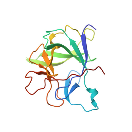
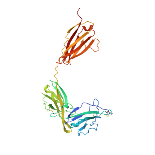
TYPE-1 INTERLEUKIN-1 RECEPTOR COMPLEXED WITH INTERLEUKIN-1 BETA
- PDB DOI: https://doi.org/10.2210/pdb1ITB/pdb
- Classification: COMPLEX (IMMUNOGLOBULIN/RECEPTOR)
- Organism(s): Homo sapiens
- Expression System: Escherichia coli
- Mutation(s): No
- Deposited: 1997-01-15 Released: 1998-02-04
- Deposition Author(s): Vigers, G.P.A., Anderson, L.J., Caffes, P., Brandhuber, B.J.
Experimental Data Snapshot
- Method: X-RAY DIFFRACTION
- Resolution: 2.50 Å
- R-Value Free: 0.325 (Depositor), 0.310 (DCC)
- R-Value Work: 0.229 (Depositor), 0.230 (DCC)
- R-Value Observed: 0.229 (Depositor)
wwPDB Validation 3D Report Full Report
- 👁 Image
Download Mendeley
Crystal structure of the type-I interleukin-1 receptor complexed with interleukin-1beta.
Vigers, G.P., Anderson, L.J., Caffes, P., Brandhuber, B.J.(1997) Nature 386: 190-194
- PubMed: 9062193 Search on PubMed
- DOI: https://doi.org/10.1038/386190a0
- Primary Citation Related Structures:
1ITB - PubMed Abstract:
Interleukin-1 (IL-1) is an important mediator of inflammatory disease. The IL-1 family currently consists of two agonists, IL-1alpha and IL-1beta, and one antagonist, IL-1ra. Each of these molecules binds to the type I IL-1 receptor (IL1R). The binding of IL-1alpha or IL-1beta to IL1R is an early step in IL-1 signal transduction and blocking this interaction may therefore be a useful target for the development of new drugs. Here we report the three-dimensional structure of IL-1beta bound to the extracellular domain of IL1R (s-IL1R) at 2.5 A resolution. IL-1beta binds to s-IL1R with a 1:1 stoichiometry. The crystal structure shows that s-IL1R consists of three immunoglobulin-like domains which wrap around IL-1beta in a manner distinct from the structures of previously described cytokine-receptor complexes. The two receptor-binding regions on IL-1beta identified by site-directed mutagenesis both contact the receptor: one binds to the first two domains of the receptor, while the other binds exclusively to the third domain.
- Amgen Inc., Boulder, Colorado 80301, USA.
Organizational Affiliation:
Explore in 3D: Structure | Sequence Annotations | Electron Density | Validation Report
Explore in 3D: Structure | Sequence Annotations | Electron Density | Validation Report
Global Symmetry: Asymmetric - C1
Global Stoichiometry: Hetero 2-mer - A1B1
Find Similar Assemblies
Biological assembly 1 assigned by authors.
Macromolecule Content
- Total Structure Weight: 53.62 kDa
- Atom Count: 3,751
- Modeled Residue Count: 463
- Deposited Residue Count: 468
- Unique protein chains: 2
Entity ID: 1 | |||||
|---|---|---|---|---|---|
| Molecule | Chains | Sequence Length | Organism | Details | Image |
| INTERLEUKIN-1 BETA | 153 | Homo sapiens | Mutation(s): 0 | 👁 Image | |
UniProt & NIH Common Fund Data Resources | |||||
Find proteins for P01584 (Homo sapiens) Explore P01584 Go to UniProtKB: P01584 | |||||
PHAROS: P01584 GTEx: ENSG00000125538 | |||||
Entity Groups | |||||
| Sequence Clusters | 30% Identity50% Identity70% Identity90% Identity95% Identity100% Identity | ||||
| UniProt Group | P01584 | ||||
Sequence AnnotationsExpand | |||||
| |||||
Entity ID: 2 | |||||
|---|---|---|---|---|---|
| Molecule | Chains | Sequence Length | Organism | Details | Image |
| TYPE 1 INTERLEUKIN-1 RECEPTOR | 315 | Homo sapiens | Mutation(s): 0 EC: 3.2.2.6 | 👁 Image | |
UniProt & NIH Common Fund Data Resources | |||||
Find proteins for P14778 (Homo sapiens) Explore P14778 Go to UniProtKB: P14778 | |||||
PHAROS: P14778 GTEx: ENSG00000115594 | |||||
Entity Groups | |||||
| Sequence Clusters | 30% Identity50% Identity70% Identity90% Identity95% Identity100% Identity | ||||
| UniProt Group | P14778 | ||||
Sequence AnnotationsExpand | |||||
| |||||
Experimental Data
- Method: X-RAY DIFFRACTION
- Resolution: 2.50 Å
- R-Value Free: 0.325 (Depositor), 0.310 (DCC)
- R-Value Work: 0.229 (Depositor), 0.230 (DCC)
- R-Value Observed: 0.229 (Depositor)
| Length ( Å ) | Angle ( ˚ ) |
|---|---|
| a = 146.952 | α = 90 |
| b = 68.452 | β = 108.95 |
| c = 65.867 | γ = 90 |
| Software Name | Purpose |
|---|---|
| DENZO | data reduction |
| SCALEPACK | data scaling |
| X-PLOR | model building |
| X-PLOR | refinement |
| X-PLOR | phasing |
Deposition Data
- Released Date: 1998-02-04 Deposition Author(s): Vigers, G.P.A., Anderson, L.J., Caffes, P., Brandhuber, B.J.
Revision History (Full details and data files)
- Version 1.0: 1998-02-04
Type: Initial release - Version 1.1: 2008-03-24
Changes: Version format compliance - Version 1.2: 2011-07-13
Changes: Version format compliance - Version 1.3: 2024-10-23
Changes: Data collection, Database references, Structure summary