
- FASTA Sequence
- PDBx/mmCIF Format
- PDBx/mmCIF Format (gz)
- BinaryCIF Format (gz)
- Legacy PDB Format
- Legacy PDB Format (gz)
- PDBML/XML Format (gz)
- Structure Factors (CIF)
- Structure Factors (CIF - gz)
- Validation Full (PDF - gz)
- Validation (XML - gz)
- Validation (CIF - gz)
- Validation 2fo-fc coefficients (CIF - gz)
- Validation fo-fc coefficients (CIF - gz)
- Biological Assembly 1 (CIF - gz)
- Biological Assembly 1 (PDB - gz)
1L2H | pdb_00001l2h
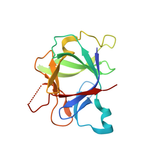
Crystal structure of Interleukin 1-beta F42W/W120F mutant
- PDB DOI: https://doi.org/10.2210/pdb1L2H/pdb
- Classification: IMMUNE SYSTEM
- Organism(s): Homo sapiens
- Expression System: Escherichia coli
- Mutation(s): Yes
- Deposited: 2002-02-21 Released: 2003-02-04
- Deposition Author(s): Rudolph, M.G., Kelker, M.S., Schneider, T.R., Yeates, T.O., Oseroff, V., Heidary, D.K., Jennings, P.A., Wilson, I.A.
Experimental Data Snapshot
- Method: X-RAY DIFFRACTION
- Resolution: 1.54 Å
- R-Value Free: 0.193 (Depositor), 0.190 (DCC)
- R-Value Work: 0.154 (Depositor), 0.150 (DCC)
wwPDB Validation 3D Report Full Report
- 👁 Image
Download Mendeley
Use of multiple anomalous dispersion to phase highly merohedrally twinned crystals of interleukin-1beta.
Rudolph, M.G., Kelker, M.S., Schneider, T.R., Yeates, T.O., Oseroff, V., Heidary, D.K., Jennings, P.A., Wilson, I.A.(2003) Acta Crystallogr D Biol Crystallogr 59: 290-298
- PubMed: 12554939 Search on PubMed
- DOI: https://doi.org/10.1107/s0907444902021704
- Primary Citation Related Structures:
1L2H - PubMed Abstract:
The crystal structure at 1.54 A resolution of a double mutant of interleukin-1beta (F42W/W120F), a cytokine secreted by macrophages, was determined by multiple-wavelength anomalous dispersion (MAD) using data from highly twinned selenomethionine-modified crystals. The space group is P4(3), with unit-cell parameters a = b = 53.9, c = 77.4 A. Self-rotation function analysis and various intensity statistics revealed the presence of merohedral twinning in crystals of both the native (twinning fraction alpha approximately 0.35) and SeMet (alpha approximately 0.40) forms. Structure determination and refinement are discussed with emphasis on the possible reasons for successful phasing using untreated twinned MAD data.
- Department of Molecular Biology and The Skaggs Institute for Chemical Biology, The Scripps Research Institute, La Jolla, CA 92037, USA.
Organizational Affiliation:
Explore in 3D: Structure | Sequence Annotations | Electron Density | Validation Report
Explore in 3D: Structure | Sequence Annotations | Electron Density | Validation Report
Global Symmetry: Asymmetric - C1
Global Stoichiometry: Monomer - A1
Find Similar Assemblies
Biological assembly 1 assigned by authors.
Macromolecule Content
- Total Structure Weight: 17.4 kDa
- Atom Count: 1,294
- Modeled Residue Count: 144
- Deposited Residue Count: 153
- Unique protein chains: 1
Entity ID: 1 | |||||
|---|---|---|---|---|---|
| Molecule | Chains | Sequence Length | Organism | Details | Image |
| Interleukin 1-beta | 153 | Homo sapiens | Mutation(s): 2 | 👁 Image | |
UniProt & NIH Common Fund Data Resources | |||||
Find proteins for P01584 (Homo sapiens) Explore P01584 Go to UniProtKB: P01584 | |||||
PHAROS: P01584 GTEx: ENSG00000125538 | |||||
Entity Groups | |||||
| Sequence Clusters | 30% Identity50% Identity70% Identity90% Identity95% Identity100% Identity | ||||
| UniProt Group | P01584 | ||||
Sequence AnnotationsExpand | |||||
| |||||
Experimental Data
- Method: X-RAY DIFFRACTION
- Resolution: 1.54 Å
- R-Value Free: 0.193 (Depositor), 0.190 (DCC)
- R-Value Work: 0.154 (Depositor), 0.150 (DCC)
| Length ( Å ) | Angle ( ˚ ) |
|---|---|
| a = 53.89 | α = 90 |
| b = 53.89 | β = 90 |
| c = 77.36 | γ = 90 |
| Software Name | Purpose |
|---|---|
| DENZO | data reduction |
| SCALEPACK | data scaling |
| SOLVE | phasing |
| SHELXL-97 | refinement |
Deposition Data
- Released Date: 2003-02-04 Deposition Author(s): Rudolph, M.G., Kelker, M.S., Schneider, T.R., Yeates, T.O., Oseroff, V., Heidary, D.K., Jennings, P.A., Wilson, I.A.
Revision History (Full details and data files)
- Version 1.0: 2003-02-04
Type: Initial release - Version 1.1: 2008-04-28
Changes: Version format compliance - Version 1.2: 2011-07-13
Changes: Version format compliance - Version 1.3: 2021-10-27
Changes: Database references - Version 1.4: 2024-02-14
Changes: Data collection