
- FASTA Sequence
- PDBx/mmCIF Format
- PDBx/mmCIF Format (gz)
- BinaryCIF Format (gz)
- Legacy PDB Format
- Legacy PDB Format (gz)
- PDBML/XML Format (gz)
- NMR Restraints (Text)
- NMR Restraints (Text - gz)
- NMR Restraints v2 (Text)
- NMR Restraints v2 (Text - gz)
- Validation Full (PDF - gz)
- Validation (XML - gz)
- Validation (CIF - gz)
- Biological Assembly 1 (CIF - gz)
- Biological Assembly 1 (PDB - gz)
1MDI | pdb_00001mdi
HIGH RESOLUTION SOLUTION NMR STRUCTURE OF MIXED DISULFIDE INTERMEDIATE BETWEEN MUTANT HUMAN THIOREDOXIN AND A 13 RESIDUE PEPTIDE COMPRISING ITS TARGET SITE IN HUMAN NFKB
- PDB DOI: https://doi.org/10.2210/pdb1MDI/pdb
- Classification: COMPLEX (ELECTRON TRANSPORT/PEPTIDE)
- Organism(s): Homo sapiens
- Mutation(s): No
- Deposited: 1995-02-27 Released: 1995-06-03
- Deposition Author(s): Clore, G.M., Qin, J., Gronenborn, A.M.
Experimental Data Snapshot
- Method: SOLUTION NMR
- Conformers Submitted: 1
wwPDB Validation 3D Report Full Report
- 👁 Image
Download Mendeley
Solution structure of human thioredoxin in a mixed disulfide intermediate complex with its target peptide from the transcription factor NF kappa B.
Qin, J., Clore, G.M., Kennedy, W.M., Huth, J.R., Gronenborn, A.M.(1995) Structure 3: 289-297
- PubMed: 7788295 Search on PubMed
- DOI: https://doi.org/10.1016/s0969-2126(01)00159-9
- Primary Citation Related Structures:
1MDI, 1MDJ, 1MDK - PubMed Abstract:
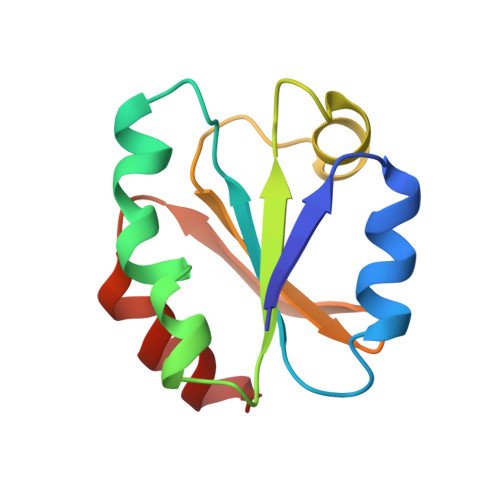
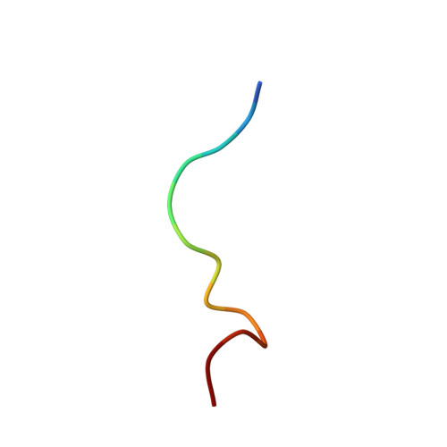
Human thioredoxin is a 12 kDa cellular redox protein that plays a key role in maintaining the redox environment of the cell. It has recently been shown to be responsible for activating the DNA-binding properties of the cellular transcription factor, NF kappa B, by reducing a disulfide bond involving Cys62 of the p50 subunit. Using multidimensional heteronuclear-edited and hetero-nuclear-filtered NMR spectroscopy, we have solved the solution structure of a complex of human thioredoxin and a 13-residue peptide extending from residues 56-68 of p50, representing a kinetically stable mixed disulfide intermediate along the reaction pathway. The NF kappa B peptide is located in a long boot-shaped cleft on the surface of human thioredoxin delineated by the active-site loop, helices alpha 2, alpha 3 and alpha 4, and strands beta 3 and beta 4. The peptide adopts a crescent-like conformation with a smooth 110 degrees bend centered around residue 60 which permits it to follow the path of the cleft. In addition to the intermolecular disulfide bridge between Cys32 of human thioredoxin and Cys62 of the peptide, the complex is stabilized by numerous hydrogen-bonding, electrostatic and hydrophobic interactions which involve residues 57-65 of the NF kappa B peptide and confer substrate specificity. These structural features permit one to suggest the specificity requirements for human thioredoxin-catalyzed disulfide bond reduction of proteins.
- Laboratory of Chemical Physics, National Institute of Diabetes and Digestive and Kidney Diseases, National Institutes of Health, Bethesda, MD 20892-0520, USA.
Organizational Affiliation:
Explore in 3D: Structure | Sequence Annotations | Validation Report
Global Symmetry: Asymmetric - C1
Global Stoichiometry: Hetero 2-mer - A1B1
Find Similar Assemblies
Biological assembly 1 assigned by authors.
Macromolecule Content
- Total Structure Weight: 13.15 kDa
- Atom Count: 932
- Modeled Residue Count: 118
- Deposited Residue Count: 118
- Unique protein chains: 2
Entity ID: 1 | |||||
|---|---|---|---|---|---|
| Molecule | Chains | Sequence Length | Organism | Details | Image |
| THIOREDOXIN | 105 | Homo sapiens | Mutation(s): 0 | 👁 Image | |
UniProt & NIH Common Fund Data Resources | |||||
Find proteins for P10599 (Homo sapiens) Explore P10599 Go to UniProtKB: P10599 | |||||
PHAROS: P10599 GTEx: ENSG00000136810 | |||||
Entity Groups | |||||
| Sequence Clusters | 30% Identity50% Identity70% Identity90% Identity95% Identity100% Identity | ||||
| UniProt Group | P10599 | ||||
Sequence AnnotationsExpand | |||||
| |||||
Find similar proteins by: Sequence | 3D Structure
Entity ID: 2 | |||||
|---|---|---|---|---|---|
| Molecule | Chains | Sequence Length | Organism | Details | Image |
| TARGET SITE IN HUMAN NFKB | 13 | Homo sapiens | Mutation(s): 0 Gene Names: NFKB1 | 👁 Image | |
UniProt & NIH Common Fund Data Resources | |||||
Find proteins for P19838 (Homo sapiens) Explore P19838 Go to UniProtKB: P19838 | |||||
PHAROS: P19838 GTEx: ENSG00000109320 | |||||
Entity Groups | |||||
| Sequence Clusters | 30% Identity50% Identity70% Identity90% Identity95% Identity100% Identity | ||||
| UniProt Group | P19838 | ||||
Sequence AnnotationsExpand | |||||
| |||||
Experimental Data
- Method: SOLUTION NMR
- Conformers Submitted: 1
Deposition Data
- Released Date: 1995-06-03 Deposition Author(s): Clore, G.M., Qin, J., Gronenborn, A.M.
Revision History (Full details and data files)
- Version 1.0: 1995-06-03
Type: Initial release - Version 1.1: 2008-03-24
Changes: Version format compliance - Version 1.2: 2011-07-13
Changes: Version format compliance - Version 1.3: 2022-02-23
Changes: Database references, Derived calculations, Other - Version 1.4: 2024-10-16
Changes: Data collection, Structure summary