
- FASTA Sequence
- PDBx/mmCIF Format
- PDBx/mmCIF Format (gz)
- BinaryCIF Format (gz)
- Legacy PDB Format
- Legacy PDB Format (gz)
- PDBML/XML Format (gz)
- Structure Factors (CIF)
- Structure Factors (CIF - gz)
- Validation Full (PDF - gz)
- Validation (XML - gz)
- Validation (CIF - gz)
- Validation 2fo-fc coefficients (CIF - gz)
- Validation fo-fc coefficients (CIF - gz)
- Biological Assembly 1 (CIF - gz)
- Biological Assembly 1 (PDB - gz)
1PIN | pdb_00001pin
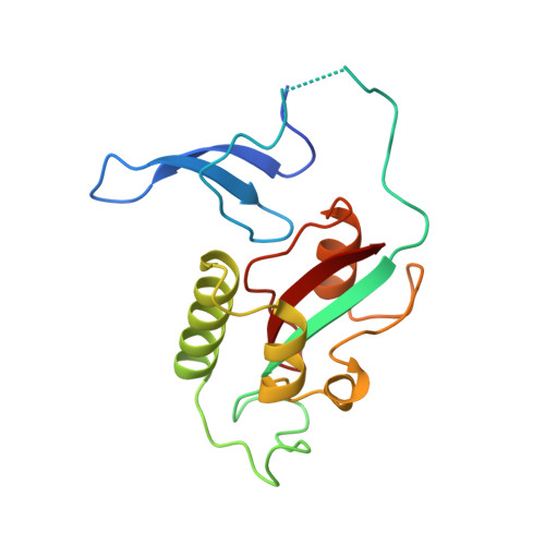
PIN1 PEPTIDYL-PROLYL CIS-TRANS ISOMERASE FROM HOMO SAPIENS
- PDB DOI: https://doi.org/10.2210/pdb1PIN/pdb
- Classification: ISOMERASE
- Organism(s): Homo sapiens
- Expression System: Escherichia coli BL21(DE3)
- Mutation(s): No
- Deposited: 1998-06-21 Released: 1998-10-14
- Deposition Author(s): Noel, J.P., Ranganathan, R., Hunter, T.
Experimental Data Snapshot
- Method: X-RAY DIFFRACTION
- Resolution: 1.35 Å
- R-Value Free: 0.266 (Depositor), 0.400 (DCC)
- R-Value Work: 0.223 (Depositor), 0.380 (DCC)
- R-Value Observed: 0.223 (Depositor)
wwPDB Validation 3D Report Full Report
- 👁 Image
Download Mendeley
Structural and functional analysis of the mitotic rotamase Pin1 suggests substrate recognition is phosphorylation dependent.
Ranganathan, R., Lu, K.P., Hunter, T., Noel, J.P.(1997) Cell 89: 875-886
- PubMed: 9200606 Search on PubMed
- DOI: https://doi.org/10.1016/s0092-8674(00)80273-1
- Primary Citation Related Structures:
1PIN - PubMed Abstract:
The human rotamase or peptidyl-prolyl cis-trans isomerase Pin1 is a conserved mitotic regulator essential for the G2/M transition of the eukaryotic cell cycle. We report the 1.35 A crystal structure of Pin1 complexed with an AlaPro dipeptide and the initial characterization of Pin1's functional properties. The crystallographic structure as well as pH titration studies and mutagenesis of an active site cysteine suggest a catalytic mechanism that includes general acid-base and covalent catalysis during peptide bond isomerization. Pin1 displays a preference for an acidic residue N-terminal to the isomerized proline bond due to interaction of this acidic side chain with a basic cluster. This raises the possibility of phosphorylation-mediated control of Pin1-substrate interactions in cell cycle regulation.
- Structural Biology Laboratory, The Salk Institute for Biological Studies, La Jolla, California 92037, USA.
Organizational Affiliation:
Explore in 3D: Structure | Sequence Annotations | Electron Density | Validation Report | Ligand Interaction (1PG)
Explore in 3D: Structure | Sequence Annotations | Electron Density | Validation Report | Ligand Interaction (1PG)
Global Symmetry: Cyclic - C2 (Explore in 3D)
Global Stoichiometry: Homo 2-mer - A2
Find Similar Assemblies
Biological assembly 1 assigned by authors.
Macromolecule Content
- Total Structure Weight: 19.08 kDa
- Atom Count: 1,458
- Modeled Residue Count: 153
- Deposited Residue Count: 163
- Unique protein chains: 1
Entity ID: 1 | |||||
|---|---|---|---|---|---|
| Molecule | Chains | Sequence Length | Organism | Details | Image |
| PEPTIDYL-PROLYL CIS-TRANS ISOMERASE | 163 | Homo sapiens | Mutation(s): 0 Gene Names: PIN1 EC: 5.2.1.8 | 👁 Image | |
UniProt & NIH Common Fund Data Resources | |||||
Find proteins for Q13526 (Homo sapiens) Explore Q13526 Go to UniProtKB: Q13526 | |||||
PHAROS: Q13526 GTEx: ENSG00000127445 | |||||
Entity Groups | |||||
| Sequence Clusters | 30% Identity50% Identity70% Identity90% Identity95% Identity100% Identity | ||||
| UniProt Group | Q13526 | ||||
Sequence AnnotationsExpand | |||||
| |||||
| Ligands 4 Unique | |||||
|---|---|---|---|---|---|
| ID | Chains | Name / Formula / InChI Key | 2D Diagram | 3D Interactions | |
| 1PG Query on 1PG Download Ideal Coordinates CCD File | E [auth A], F [auth A] | 2-(2-{2-[2-(2-METHOXY-ETHOXY)-ETHOXY]-ETHOXY}-ETHOXY)-ETHANOL C11 H24 O6 SLNYBUIEAMRFSZ-UHFFFAOYSA-N | 👁 Image | ||
| PRO Query on PRO Download Ideal Coordinates CCD File | C [auth A] | PROLINE C5 H9 N O2 ONIBWKKTOPOVIA-BYPYZUCNSA-N | 👁 Image | ||
| SO4 Query on SO4 Download Ideal Coordinates CCD File | D [auth A] | SULFATE ION O4 S QAOWNCQODCNURD-UHFFFAOYSA-L | 👁 Image | ||
| ALA Query on ALA Download Ideal Coordinates CCD File | B [auth A] | ALANINE C3 H7 N O2 QNAYBMKLOCPYGJ-REOHCLBHSA-N | 👁 Image | ||
Experimental Data
- Method: X-RAY DIFFRACTION
- Resolution: 1.35 Å
- R-Value Free: 0.266 (Depositor), 0.400 (DCC)
- R-Value Work: 0.223 (Depositor), 0.380 (DCC)
- R-Value Observed: 0.223 (Depositor)
| Length ( Å ) | Angle ( ˚ ) |
|---|---|
| a = 49 | α = 90 |
| b = 49 | β = 90 |
| c = 137.8 | γ = 90 |
| Software Name | Purpose |
|---|---|
| X-PLOR | model building |
| X-PLOR | refinement |
| DENZO | data reduction |
| SCALEPACK | data scaling |
| X-PLOR | phasing |
Deposition Data
- Released Date: 1998-10-14 Deposition Author(s): Noel, J.P., Ranganathan, R., Hunter, T.
Revision History (Full details and data files)
- Version 1.0: 1998-10-14
Type: Initial release - Version 1.1: 2008-03-24
Changes: Version format compliance - Version 1.2: 2011-07-13
Changes: Version format compliance - Version 1.3: 2013-04-24
Changes: Other - Version 1.4: 2014-02-26
Changes: Other - Version 2.0: 2023-07-26
Type: Remediation
Changes: Advisory, Atomic model, Data collection, Database references, Derived calculations, Other - Version 2.1: 2024-05-22
Changes: Data collection