
1SVC | pdb_00001svc
NFKB P50 HOMODIMER BOUND TO DNA
- PDB DOI: https://doi.org/10.2210/pdb1SVC/pdb
- NAKB: 1SVC
- Classification: TRANSCRIPTION/DNA
- Organism(s): Homo sapiens
- Mutation(s): Yes
- Deposited: 1995-11-27 Released: 1996-06-10
- Deposition Author(s): Mueller, C.W., Harrison, S.C.
Experimental Data Snapshot
- Method: X-RAY DIFFRACTION
- Resolution: 2.60 Å
- R-Value Free: 0.286 (Depositor)
- R-Value Work: 0.225 (Depositor)
- R-Value Observed: 0.225 (Depositor)
wwPDB Validation 3D Report Full Report
- 👁 Image
Download Mendeley
Structure of the NF-kappa B p50 homodimer bound to DNA.
Muller, C.W., Rey, F.A., Sodeoka, M., Verdine, G.L., Harrison, S.C.(1995) Nature 373: 311-317
- PubMed: 7830764 Search on PubMed
- DOI: https://doi.org/10.1038/373311a0
- Primary Citation Related Structures:
1SVC - PubMed Abstract:
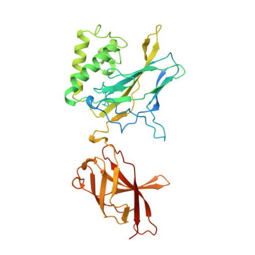
The structure of a large fragment of the p50 subunit of the human transcription factor NF-kappa B, bound as a homodimer to DNA, reveals that the Rel-homology region has two beta-barrel domains that grip DNA in the major groove. Both domains contact the DNA backbone. The amino-terminal specificity domain contains a recognition loop that interacts with DNA bases; the carboxy-terminal dimerization domain bears the site of I-kappa B interaction. The folds of these domains are related to immunoglobulin-like modules. The amino-terminal domain also resembles the core domain of p53.
- Howard Hughes Medical Institute, Cambridge, Massachusetts 02138.
Organizational Affiliation:
Explore in 3D: Structure | Sequence Annotations | Validation Report
Global Symmetry: Cyclic - C2 (Explore in 3D)
Global Stoichiometry: Homo 2-mer - A2
Find Similar Assemblies
Biological assembly 1 assigned by authors.
Macromolecule Content
- Total Structure Weight: 47.01 kDa
- Atom Count: 2,903
- Modeled Residue Count: 330
- Deposited Residue Count: 384
- Unique protein chains: 1
- Unique nucleic acid chains: 1
Entity ID: 2 | |||||
|---|---|---|---|---|---|
| Molecule | Chains | Sequence Length | Organism | Details | Image |
| PROTEIN (NUCLEAR FACTOR KAPPA-B (NF-KB)) | B [auth P] | 365 | Homo sapiens | Mutation(s): 1 | 👁 Image |
UniProt & NIH Common Fund Data Resources | |||||
Find proteins for P19838 (Homo sapiens) Explore P19838 Go to UniProtKB: P19838 | |||||
PHAROS: P19838 GTEx: ENSG00000109320 | |||||
Entity Groups | |||||
| Sequence Clusters | 30% Identity50% Identity70% Identity90% Identity95% Identity100% Identity | ||||
| UniProt Group | P19838 | ||||
Sequence AnnotationsExpand | |||||
| |||||
Find similar nucleic acids by: Sequence
Entity ID: 1 | |||||
|---|---|---|---|---|---|
| Molecule | Chains | Length | Organism | Image | |
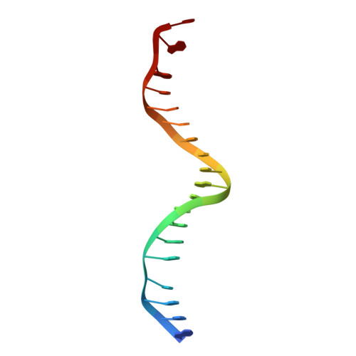
| DNA (5'-D(*AP*GP*AP*TP*GP*GP*GP*GP*AP*AP*TP*CP*CP*CP*CP*TP*A P*GP*A)-3') | A [auth D] | 19 | N/A | 👁 Image | |
Sequence AnnotationsExpand | |||||
| |||||
Experimental Data
- Method: X-RAY DIFFRACTION
- Resolution: 2.60 Å
- R-Value Free: 0.286 (Depositor)
- R-Value Work: 0.225 (Depositor)
- R-Value Observed: 0.225 (Depositor)
| Length ( Å ) | Angle ( ˚ ) |
|---|---|
| a = 137 | α = 90 |
| b = 137 | β = 90 |
| c = 57 | γ = 90 |
| Software Name | Purpose |
|---|---|
| X-PLOR | refinement |
Deposition Data
- Released Date: 1996-06-10 Deposition Author(s): Mueller, C.W., Harrison, S.C.
Revision History (Full details and data files)
- Version 1.0: 1996-06-10
Type: Initial release - Version 1.1: 2008-05-22
Changes: Version format compliance - Version 1.2: 2011-07-13
Changes: Version format compliance - Version 1.3: 2021-11-03
Changes: Database references - Version 1.4: 2024-02-14
Changes: Data collection