
- FASTA Sequence
- PDBx/mmCIF Format
- PDBx/mmCIF Format (gz)
- BinaryCIF Format (gz)
- Legacy PDB Format
- Legacy PDB Format (gz)
- PDBML/XML Format (gz)
- Structure Factors (CIF)
- Structure Factors (CIF - gz)
- Validation Full (PDF - gz)
- Validation (XML - gz)
- Validation (CIF - gz)
- Validation 2fo-fc coefficients (CIF - gz)
- Validation fo-fc coefficients (CIF - gz)
- Biological Assembly 1 (CIF - gz)
- Biological Assembly 1 (PDB - gz)
2AO6 | pdb_00002ao6
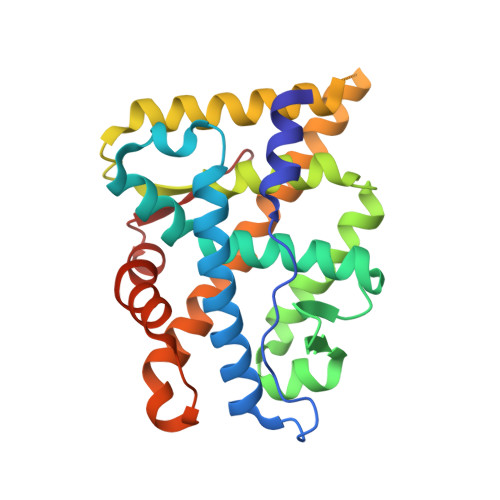
Crystal structure of the human androgen receptor ligand binding domain bound with TIF2(iii) 740-753 peptide and R1881
- PDB DOI: https://doi.org/10.2210/pdb2AO6/pdb
- Entry: 2AO6 supersedes: 1XQ2
- Classification: TRANSCRIPTION
- Organism(s): Homo sapiens
- Expression System: Escherichia coli BL21(DE3)
- Mutation(s): No
- Deposited: 2005-08-12 Released: 2005-08-30
- Deposition Author(s): He, B., Gampe Jr., R.T., Kole, A.J., Hnat, A.T., Stanley, T.B., An, G., Stewart, E.L., Kalman, R.I., Minges, J.T., Wilson, E.M.
Experimental Data Snapshot
- Method: X-RAY DIFFRACTION
- Resolution: 1.89 Å
- R-Value Free: 0.242 (Depositor)
- R-Value Work: 0.220 (Depositor), 0.220 (DCC)
Starting Model: experimental
View more details
- 👁 Image
Download Mendeley
Structural basis for androgen receptor interdomain and coactivator interactions suggests a transition in nuclear receptor activation function dominance
He, B., Gampe Jr., R.T., Kole, A.J., Hnat, A.T., Stanley, T.B., An, G., Stewart, E.L., Kalman, R.I., Minges, J.T., Wilson, E.M.(2004) Mol Cell 16: 425-438
- PubMed: 15525515 Search on PubMed
- DOI: https://doi.org/10.1016/j.molcel.2004.09.036
- Primary Citation Related Structures:
1XOW, 1XQ3, 2AO6 - PubMed Abstract:
The androgen receptor (AR) is required for male sex development and contributes to prostate cancer cell survival. In contrast to other nuclear receptors that bind the LXXLL motifs of coactivators, the AR ligand binding domain is preferentially engaged in an interdomain interaction with the AR FXXLF motif. Reported here are crystal structures of the ligand-activated AR ligand binding domain with and without bound FXXLF and LXXLL peptides. Key residues that establish motif binding specificity are identified through comparative structure-function and mutagenesis studies. A mechanism in prostate cancer is suggested by a functional AR mutation at a specificity-determining residue that recovers coactivator LXXLL motif binding. An activation function transition hypothesis is proposed in which an evolutionary decline in LXXLL motif binding parallels expansion and functional dominance of the NH(2)-terminal transactivation domain in the steroid receptor subfamily.
- Laboratories for Reproductive Biology, University of North Carolina at Chapel Hill, Chapel Hill, NC 27599, USA.
Organizational Affiliation:
Explore in 3D: Structure | Sequence Annotations | Electron Density | Validation Report | Ligand Interaction (R18)
Explore in 3D: Structure | Sequence Annotations | Electron Density | Validation Report | Ligand Interaction (R18)
Global Symmetry: Asymmetric - C1
Global Stoichiometry: Hetero 2-mer - A1B1
Find Similar Assemblies
Biological assembly 1 assigned by authors and generated by PISA (software)
Macromolecule Content
- Total Structure Weight: 32.14 kDa
- Atom Count: 2,186
- Modeled Residue Count: 253
- Deposited Residue Count: 274
- Unique protein chains: 2
Entity ID: 1 | |||||
|---|---|---|---|---|---|
| Molecule | Chains | Sequence Length | Organism | Details | Image |
| androgen receptor | 249 | Homo sapiens | Mutation(s): 0 Gene Names: Ar, Nr3c4 | 👁 Image | |
UniProt & NIH Common Fund Data Resources | |||||
Find proteins for P10275 (Homo sapiens) Explore P10275 Go to UniProtKB: P10275 | |||||
PHAROS: P10275 GTEx: ENSG00000169083 | |||||
Entity Groups | |||||
| Sequence Clusters | 30% Identity50% Identity70% Identity90% Identity95% Identity100% Identity | ||||
| UniProt Group | P10275 | ||||
Sequence AnnotationsExpand | |||||
| |||||
Find similar proteins by: Sequence | 3D Structure
Entity ID: 2 | |||||
|---|---|---|---|---|---|
| Molecule | Chains | Sequence Length | Organism | Details | Image |
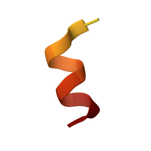
| 14-mer fragment of Nuclear receptor coactivator 2 | 25 | N/A | Mutation(s): 0 | 👁 Image | |
UniProt & NIH Common Fund Data Resources | |||||
Find proteins for Q15596 (Homo sapiens) Explore Q15596 Go to UniProtKB: Q15596 | |||||
PHAROS: Q15596 GTEx: ENSG00000140396 | |||||
Entity Groups | |||||
| Sequence Clusters | 30% Identity50% Identity70% Identity90% Identity95% Identity100% Identity | ||||
| UniProt Group | Q15596 | ||||
Sequence AnnotationsExpand | |||||
| |||||
| Ligands 1 Unique | |||||
|---|---|---|---|---|---|
| ID | Chains | Name / Formula / InChI Key | 2D Diagram | 3D Interactions | |
| R18 Query on R18 Download Ideal Coordinates CCD File | C [auth A] | (17BETA)-17-HYDROXY-17-METHYLESTRA-4,9,11-TRIEN-3-ONE C19 H24 O2 CCCIJQPRIXGQOE-XWSJACJDSA-N | 👁 Image | ||
Experimental Data
- Method: X-RAY DIFFRACTION
- Resolution: 1.89 Å
- R-Value Free: 0.242 (Depositor)
- R-Value Work: 0.220 (Depositor), 0.220 (DCC)
| Length ( Å ) | Angle ( ˚ ) |
|---|---|
| a = 54.58 | α = 90 |
| b = 66.7 | β = 90 |
| c = 69.4 | γ = 90 |
| Software Name | Purpose |
|---|---|
| MAR345 | data collection |
| HKL-2000 | data reduction |
| CNX | refinement |
| HKL-2000 | data scaling |
| CNX | phasing |
Deposition Data
- Released Date: 2005-08-30 Deposition Author(s): He, B., Gampe Jr., R.T., Kole, A.J., Hnat, A.T., Stanley, T.B., An, G., Stewart, E.L., Kalman, R.I., Minges, J.T., Wilson, E.M.
- This entry supersedes: 1XQ2
Revision History (Full details and data files)
- Version 1.0: 2005-08-30
Type: Initial release - Version 1.1: 2008-04-30
Changes: Version format compliance - Version 1.2: 2011-07-13
Changes: Version format compliance - Version 1.3: 2017-10-11
Changes: Refinement description - Version 1.4: 2023-08-23
Changes: Data collection, Database references, Derived calculations, Refinement description