
- FASTA Sequence
- PDBx/mmCIF Format
- PDBx/mmCIF Format (gz)
- BinaryCIF Format (gz)
- Legacy PDB Format
- Legacy PDB Format (gz)
- PDBML/XML Format (gz)
- Structure Factors (CIF)
- Structure Factors (CIF - gz)
- Validation Full (PDF - gz)
- Validation (XML - gz)
- Validation (CIF - gz)
- Validation 2fo-fc coefficients (CIF - gz)
- Validation fo-fc coefficients (CIF - gz)
- Biological Assembly 1 (CIF - gz)
- Biological Assembly 1 (PDB - gz)
2ATO | pdb_00002ato
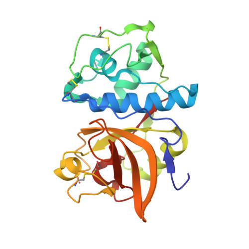
Crystal structure of Human Cathepsin K in complex with myocrisin
- PDB DOI: https://doi.org/10.2210/pdb2ATO/pdb
- Classification: HYDROLASE
- Organism(s): Homo sapiens
- Expression System: Komagataella pastoris
- Mutation(s): No
- Deposited: 2005-08-25 Released: 2006-08-29
- Deposition Author(s): Weidauer, E., Yasuda, Y., Biswal, B.K., Kerr, L.D., Cherney, M.M., Gordon, R.E., James, M.N.G., Bromme, D.
Experimental Data Snapshot
- Method: X-RAY DIFFRACTION
- Resolution: 2.00 Å
- R-Value Free: 0.198 (Depositor), 0.194 (DCC)
- R-Value Work: 0.197 (Depositor), 0.192 (DCC)
- R-Value Observed: 0.197 (Depositor)
Starting Model: experimental
View more details
Literature
- 👁 Image
Download Mendeley
Effects of disease-modifying anti-rheumatic drugs (DMARDs) on the activities of rheumatoid arthritis-associated cathepsins K and S.
Weidauer, E., Yasuda, Y., Biswal, B.K., Cherny, M., James, M.N., Bromme, D.(2007) Biol Chem 388: 331-336
- PubMed: 17338641 Search on PubMed
- DOI: https://doi.org/10.1515/BC.2007.037
- Primary Citation Related Structures:
2ATO - PubMed Abstract:
Rheumatoid arthritis is an inflammatory and disabling joint disease affecting 0.5-1.5% of the population. Although various anti-inflammatory (NSAIDs) and disease-modifying (DMARDs) drugs are in clinical use, their precise mechanisms of action are not always defined. In this report, we discuss the effects of widely used DMARDs such as gold derivatives and chloroquine on cathepsins K and S, which have been implicated as critical mediators of inflammation and joint erosion in rheumatoid arthritis. We demonstrate that clinically potent gold derivatives inhibit cathepsins K and S in in vitro and cell-based assays. An X-ray analysis of the gold thiomalate/cathepsin K complex reveals that the inhibitor is bound to the active-site cysteine residue of the protease. Chloroquine, a lysosomotropic agent of lower clinical potency than gold derivatives, inhibits neutral pH-labile cathepsins intracellularly, but does not affect the neutral pH-stable cathepsin S. The potent inhibition of cathepsins implicated in the pathogenesis of rheumatoid arthritis by gold derivatives may explain the therapeutic efficacy of these drugs.
- Department of Human Genetics, Mount Sinai School of Medicine, New York, NY 10029, USA.
Organizational Affiliation:
Explore in 3D: Structure | Sequence Annotations | Electron Density | Validation Report | Ligand Interaction (MYQ)
Biological Assembly 1
Explore in 3D: Structure | Sequence Annotations | Electron Density | Validation Report | Ligand Interaction (MYQ)
Global Symmetry: Asymmetric - C1
Global Stoichiometry: Monomer - A1
Find Similar Assemblies
Biological assembly 1 assigned by authors.
Macromolecule Content
- Total Structure Weight: 23.97 kDa
- Atom Count: 1,827
- Modeled Residue Count: 215
- Deposited Residue Count: 215
- Unique protein chains: 1
Macromolecules
Entity ID: 1 | |||||
|---|---|---|---|---|---|
| Molecule | Chains | Sequence Length | Organism | Details | Image |
| Cathepsin K | 215 | Homo sapiens | Mutation(s): 0 Gene Names: CTSK, CTSO, CTSO2 EC: 3.4.22.38 | 👁 Image | |
UniProt & NIH Common Fund Data Resources | |||||
PHAROS: P43235 GTEx: ENSG00000143387 | |||||
Entity Groups | |||||
| Sequence Clusters | 30% Identity50% Identity70% Identity90% Identity95% Identity100% Identity | ||||
| UniProt Group | P43235 | ||||
Sequence AnnotationsExpand | |||||
Reference Sequence | |||||
Small Molecules
| Ligands 2 Unique | |||||
|---|---|---|---|---|---|
| ID | Chains | Name / Formula / InChI Key | 2D Diagram | 3D Interactions | |
| MYQ Download:Ideal Coordinates CCD File | C [auth A] | (S)-(1,2-DICARBOXYETHYLTHIO)GOLD C4 H5 Au O4 S XJHSMFDIQHVMCY-DKWTVANSSA-M | 👁 Image | ||
| SO4 Download:Ideal Coordinates CCD File | B [auth A] | SULFATE ION O4 S QAOWNCQODCNURD-UHFFFAOYSA-L | 👁 Image | ||
Experimental Data & Validation
Experimental Data
- Method: X-RAY DIFFRACTION
- Resolution: 2.00 Å
- R-Value Free: 0.198 (Depositor), 0.194 (DCC)
- R-Value Work: 0.197 (Depositor), 0.192 (DCC)
- R-Value Observed: 0.197 (Depositor)
| Length ( Å ) | Angle ( ˚ ) |
|---|---|
| a = 32.063 | α = 90 |
| b = 67.398 | β = 90 |
| c = 93.785 | γ = 90 |
| Software Name | Purpose |
|---|---|
| CNS | refinement |
| Blu-Ice | data collection |
| SCALEPACK | data scaling |
| CNS | phasing |
Entry History
Deposition Data
- Released Date: 2006-08-29 Deposition Author(s): Weidauer, E., Yasuda, Y., Biswal, B.K., Kerr, L.D., Cherney, M.M., Gordon, R.E., James, M.N.G., Bromme, D.
Revision History (Full details and data files)
- Version 1.0: 2006-08-29
Type: Initial release - Version 1.1: 2008-04-30
Changes: Version format compliance - Version 1.2: 2011-07-13
Changes: Version format compliance - Version 1.3: 2023-08-23
Changes: Data collection, Database references, Derived calculations, Refinement description - Version 1.4: 2024-10-30
Changes: Structure summary