
- FASTA Sequence
- PDBx/mmCIF Format
- PDBx/mmCIF Format (gz)
- BinaryCIF Format (gz)
- Legacy PDB Format
- Legacy PDB Format (gz)
- PDBML/XML Format (gz)
- Structure Factors (CIF)
- Structure Factors (CIF - gz)
- Validation Full (PDF - gz)
- Validation (XML - gz)
- Validation (CIF - gz)
- Validation 2fo-fc coefficients (CIF - gz)
- Validation fo-fc coefficients (CIF - gz)
- Biological Assembly 1 (CIF - gz)
- Biological Assembly 2 (CIF - gz)
- Biological Assembly 1 (PDB - gz)
- Biological Assembly 2 (PDB - gz)
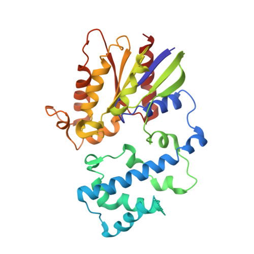
2G83 | pdb_00002g83
Structure of activated G-alpha-i1 bound to a nucleotide-state-selective peptide: Minimal determinants for recognizing the active form of a G protein alpha subunit
- PDB DOI: https://doi.org/10.2210/pdb2G83/pdb
- Classification: SIGNALING PROTEIN
- Organism(s): Homo sapiens
- Expression System: Escherichia coli BL21(DE3)
- Mutation(s): No
- Deposited: 2006-03-01 Released: 2006-10-10
- Deposition Author(s): Johnston, C.A., Ramer, J.K., Blaesius, R., Kuhlman, B., Arshavsky, V.Y., Siderovski, D.P.
Experimental Data Snapshot
- Method: X-RAY DIFFRACTION
- Resolution: 2.80 Å
- R-Value Free: 0.300 (Depositor), 0.301 (DCC)
- R-Value Work: 0.269 (Depositor), 0.271 (DCC)
- R-Value Observed: 0.301 (Depositor)
Starting Model: experimental
View more details
- 👁 Image
Download Mendeley
Minimal Determinants for Binding Activated Galpha from the Structure of a Galpha(i1)-Peptide Dimer.
Johnston, C.A., Lobanova, E.S., Shavkunov, A.S., Low, J., Ramer, J.K., Blaesius, R., Fredericks, Z., Willard, F.S., Kuhlman, B., Arshavsky, V.Y., Siderovski, D.P.(2006) Biochemistry 45: 11390-11400
- PubMed: 16981699 Search on PubMedSearch on PubMed Central
- DOI: https://doi.org/10.1021/bi0613832
- Primary Citation Related Structures:
2G83 - PubMed Abstract:
G-proteins cycle between an inactive GDP-bound state and an active GTP-bound state, serving as molecular switches that coordinate cellular signaling. We recently used phage display to identify a series of peptides that bind G alpha subunits in a nucleotide-dependent manner [Johnston, C. A., Willard, F. S., Jezyk, M. R., Fredericks, Z., Bodor, E. T., Jones, M. B., Blaesius, R., Watts, V. J., Harden, T. K., Sondek, J., Ramer, J. K., and Siderovski, D. P. (2005) Structure 13, 1069-1080]. Here we describe the structural features and functions of KB-1753, a peptide that binds selectively to GDP x AlF4(-)- and GTPgammaS-bound states of G alpha(i) subunits. KB-1753 blocks interaction of G alpha(transducin) with its effector, cGMP phosphodiesterase, and inhibits transducin-mediated activation of cGMP degradation. Additionally, KB-1753 interferes with RGS protein binding and resultant GAP activity. A fluorescent KB-1753 variant was found to act as a sensor for activated G alpha in vitro. The crystal structure of KB-1753 bound to G alpha(i1) x GDP x AlF4(-) reveals binding to a conserved hydrophobic groove between switch II and alpha3 helices and, along with supporting biochemical data and previous structural analyses, supports the notion that this is the site of effector interactions for G alpha(i) subunits.
- Department of Pharmacology, University of North Carolina School of Medicine, Chapel Hill, North Carolina 27599-7365, USA.
Organizational Affiliation:
Explore in 3D: Structure | Sequence Annotations | Electron Density | Validation Report | Ligand Interaction (GDP)
Explore in 3D: Structure | Sequence Annotations | Electron Density | Validation Report | Ligand Interaction (GDP)
Global Symmetry: Asymmetric - C1
Global Stoichiometry: Hetero 2-mer - A1B1
Find Similar Assemblies
Biological assembly 1 assigned by authors.
Explore in 3D: Structure | Sequence Annotations | Electron Density | Validation Report | Ligand Interaction (GDP)
Global Symmetry: Asymmetric - C1
Global Stoichiometry: Hetero 2-mer - A1B1
Find Similar Assemblies
Biological assembly 2 assigned by authors and generated by PISA (software)
Macromolecule Content
- Total Structure Weight: 75.55 kDa
- Atom Count: 5,190
- Modeled Residue Count: 628
- Deposited Residue Count: 648
- Unique protein chains: 2
Entity ID: 1 | |||||
|---|---|---|---|---|---|
| Molecule | Chains | Sequence Length | Organism | Details | Image |
| Guanine nucleotide-binding protein G(i), alpha-1 subunit | 313 | Homo sapiens | Mutation(s): 0 Gene Names: GNAI1 EC: 3.6.5 | 👁 Image | |
UniProt & NIH Common Fund Data Resources | |||||
Find proteins for P63096 (Homo sapiens) Explore P63096 Go to UniProtKB: P63096 | |||||
PHAROS: P63096 GTEx: ENSG00000127955 | |||||
Entity Groups | |||||
| Sequence Clusters | 30% Identity50% Identity70% Identity90% Identity95% Identity100% Identity | ||||
| UniProt Group | P63096 | ||||
Sequence AnnotationsExpand | |||||
| |||||
Find similar proteins by: Sequence | 3D Structure
Entity ID: 2 | |||||
|---|---|---|---|---|---|
| Molecule | Chains | Sequence Length | Organism | Details | Image |
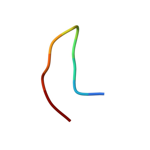
| KB-1753 phage display peptide | 11 | N/A | Mutation(s): 0 | 👁 Image | |
Entity Groups | |||||
| Sequence Clusters | 30% Identity50% Identity70% Identity90% Identity95% Identity100% Identity | ||||
Sequence AnnotationsExpand | |||||
| |||||
| Ligands 3 Unique | |||||
|---|---|---|---|---|---|
| ID | Chains | Name / Formula / InChI Key | 2D Diagram | 3D Interactions | |
| GDP Query on GDP Download Ideal Coordinates CCD File | G [auth A], J [auth B] | GUANOSINE-5'-DIPHOSPHATE C10 H15 N5 O11 P2 QGWNDRXFNXRZMB-UUOKFMHZSA-N | 👁 Image | ||
| ALF Query on ALF Download Ideal Coordinates CCD File | E [auth A], H [auth B] | TETRAFLUOROALUMINATE ION Al F4 UYOMQIYKOOHAMK-UHFFFAOYSA-J | 👁 Image | ||
| MG Query on MG Download Ideal Coordinates CCD File | F [auth A], I [auth B] | MAGNESIUM ION Mg JLVVSXFLKOJNIY-UHFFFAOYSA-N | 👁 Image | ||
Experimental Data
- Method: X-RAY DIFFRACTION
- Resolution: 2.80 Å
- R-Value Free: 0.300 (Depositor), 0.301 (DCC)
- R-Value Work: 0.269 (Depositor), 0.271 (DCC)
- R-Value Observed: 0.301 (Depositor)
| Length ( Å ) | Angle ( ˚ ) |
|---|---|
| a = 103.13 | α = 90 |
| b = 103.13 | β = 90 |
| c = 206.99 | γ = 120 |
| Software Name | Purpose |
|---|---|
| CNS | refinement |
| PDB_EXTRACT | data extraction |
| HKL-2000 | data reduction |
| HKL-2000 | data scaling |
| AMoRE | phasing |
Deposition Data
- Released Date: 2006-10-10 Deposition Author(s): Johnston, C.A., Ramer, J.K., Blaesius, R., Kuhlman, B., Arshavsky, V.Y., Siderovski, D.P.
Revision History (Full details and data files)
- Version 1.0: 2006-10-10
Type: Initial release - Version 1.1: 2008-05-01
Changes: Version format compliance - Version 1.2: 2011-07-13
Changes: Version format compliance - Version 1.3: 2023-08-30
Changes: Data collection, Database references, Derived calculations, Refinement description - Version 1.4: 2024-12-25
Changes: Advisory, Derived calculations, Structure summary