
- FASTA Sequence
- PDBx/mmCIF Format
- PDBx/mmCIF Format (gz)
- BinaryCIF Format (gz)
- Legacy PDB Format
- Legacy PDB Format (gz)
- PDBML/XML Format (gz)
- Structure Factors (CIF)
- Structure Factors (CIF - gz)
- Validation Full (PDF - gz)
- Validation (XML - gz)
- Validation (CIF - gz)
- Validation 2fo-fc coefficients (CIF - gz)
- Validation fo-fc coefficients (CIF - gz)
- Biological Assembly 1 (CIF - gz)
- Biological Assembly 1 (PDB - gz)
2H0D | pdb_00002h0d
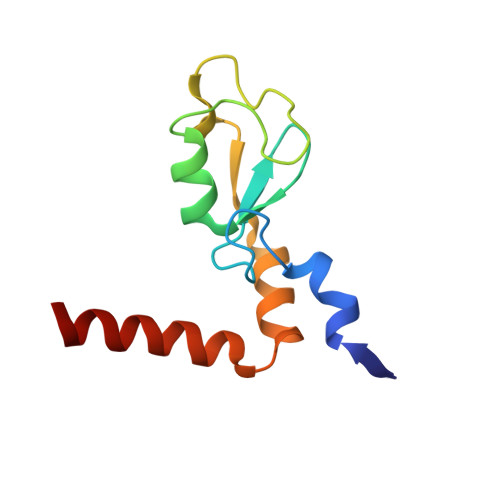
Structure of a Bmi-1-Ring1B Polycomb group ubiquitin ligase complex
- PDB DOI: https://doi.org/10.2210/pdb2H0D/pdb
- Classification: METAL BINDING PROTEIN/LIGASE
- Organism(s): Homo sapiens
- Expression System: Escherichia coli BL21(DE3)
- Mutation(s): No
- Deposited: 2006-05-14 Released: 2006-05-23
- Deposition Author(s): Xu, R.M.
Experimental Data Snapshot
- Method: X-RAY DIFFRACTION
- Resolution: 2.50 Å
- R-Value Free: 0.244 (Depositor), 0.240 (DCC)
- R-Value Work: 0.210 (Depositor), 0.210 (DCC)
- R-Value Observed: 0.211 (Depositor)
wwPDB Validation 3D Report Full Report
- 👁 Image
Download Mendeley
Structure of a Bmi-1-Ring1B Polycomb Group Ubiquitin Ligase Complex.
Li, Z., Cao, R., Wang, M., Myers, M.P., Zhang, Y., Xu, R.M.(2006) J Biological Chem 281: 20643-20649
- PubMed: 16714294 Search on PubMed
- DOI: https://doi.org/10.1074/jbc.M602461200
- Primary Citation Related Structures:
2H0D - PubMed Abstract:
Polycomb group proteins Bmi-1 and Ring1B are core subunits of the PRC1 complex, which plays important roles in the regulation of Hox gene expression, X-chromosome inactivation, tumorigenesis, and stem cell self-renewal. The RING finger protein Ring1B is an E3 ligase that participates in the ubiquitination of lysine 119 of histone H2A, and the binding of Bmi-1 stimulates the E3 ligase activity. We have mapped the regions of Bmi-1 and Ring1B required for efficient ubiquitin transfer and determined a 2.5-A structure of the Bmi-1-Ring1B core domain complex. The structure reveals that Ring1B "hugs" Bmi-1 through extensive RING domain contacts and its N-terminal tail wraps around Bmi-1. The two regions of interaction have a synergistic effect on the E3 ligase activity. Our analyses suggest a model where the Bmi-1-Ring1B complex stabilizes the interaction between the E2 enzyme and the nucleosomal substrate to allow efficient ubiquitin transfer.
- Cold Spring Harbor Laboratory, Cold Spring Harbor, New York 11724, USA.
Organizational Affiliation:
Explore in 3D: Structure | Sequence Annotations | Electron Density | Validation Report | Ligand Interaction (ZN)
Explore in 3D: Structure | Sequence Annotations | Electron Density | Validation Report | Ligand Interaction (ZN)
Global Symmetry: Asymmetric - C1
Global Stoichiometry: Hetero 2-mer - A1B1
Pseudo Symmetry: Cyclic - C2 (Explore in 3D)
Pseudo Stoichiometry: Homo 2-mer - A2
Find Similar Assemblies
Biological assembly 1 assigned by authors and generated by PISA (software)
Macromolecule Content
- Total Structure Weight: 23.11 kDa
- Atom Count: 1,624
- Modeled Residue Count: 197
- Deposited Residue Count: 197
- Unique protein chains: 2
Entity ID: 1 | |||||
|---|---|---|---|---|---|
| Molecule | Chains | Sequence Length | Organism | Details | Image |
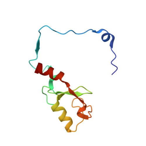
| B lymphoma Mo-MLV insertion region | 97 | Homo sapiens | Mutation(s): 0 | 👁 Image | |
UniProt & NIH Common Fund Data Resources | |||||
Find proteins for P35226 (Homo sapiens) Explore P35226 Go to UniProtKB: P35226 | |||||
PHAROS: P35226 GTEx: ENSG00000168283 | |||||
Entity Groups | |||||
| Sequence Clusters | 30% Identity50% Identity70% Identity90% Identity95% Identity100% Identity | ||||
| UniProt Group | P35226 | ||||
Sequence AnnotationsExpand | |||||
| |||||
Entity ID: 2 | |||||
|---|---|---|---|---|---|
| Molecule | Chains | Sequence Length | Organism | Details | Image |
| Ubiquitin ligase protein RING2 | 100 | Homo sapiens | Mutation(s): 0 EC: 6.3.2 (PDB Primary Data), 2.3.2.27 (UniProt) | 👁 Image | |
UniProt & NIH Common Fund Data Resources | |||||
Find proteins for Q99496 (Homo sapiens) Explore Q99496 Go to UniProtKB: Q99496 | |||||
PHAROS: Q99496 GTEx: ENSG00000121481 | |||||
Entity Groups | |||||
| Sequence Clusters | 30% Identity50% Identity70% Identity90% Identity95% Identity100% Identity | ||||
| UniProt Group | Q99496 | ||||
Sequence AnnotationsExpand | |||||
| |||||
| Ligands 1 Unique | |||||
|---|---|---|---|---|---|
| ID | Chains | Name / Formula / InChI Key | 2D Diagram | 3D Interactions | |
| ZN Query on ZN Download Ideal Coordinates CCD File
| C [auth A], D [auth A], E [auth B], F [auth B] | ZINC ION Zn PTFCDOFLOPIGGS-UHFFFAOYSA-N | 👁 Image | ||
Experimental Data
- Method: X-RAY DIFFRACTION
- Resolution: 2.50 Å
- R-Value Free: 0.244 (Depositor), 0.240 (DCC)
- R-Value Work: 0.210 (Depositor), 0.210 (DCC)
- R-Value Observed: 0.211 (Depositor)
| Length ( Å ) | Angle ( ˚ ) |
|---|---|
| a = 120.514 | α = 90 |
| b = 120.514 | β = 90 |
| c = 27.209 | γ = 120 |
| Software Name | Purpose |
|---|---|
| DENZO | data reduction |
| SCALEPACK | data scaling |
| SOLVE | phasing |
| REFMAC | refinement |
| PDB_EXTRACT | data extraction |
| MADNESS | data reduction |
Deposition Data
- Released Date: 2006-05-23 Deposition Author(s): Xu, R.M.
Revision History (Full details and data files)
- Version 1.0: 2006-05-23
Type: Initial release - Version 1.1: 2008-05-01
Changes: Version format compliance - Version 1.2: 2011-07-13
Changes: Advisory, Version format compliance - Version 1.3: 2017-10-18
Changes: Refinement description - Version 1.4: 2024-02-14
Changes: Data collection, Database references, Derived calculations