
- FASTA Sequence
- PDBx/mmCIF Format
- PDBx/mmCIF Format (gz)
- BinaryCIF Format (gz)
- Legacy PDB Format
- Legacy PDB Format (gz)
- PDBML/XML Format (gz)
- NMR Restraints (Text)
- NMR Restraints (Text - gz)
- NMR Restraints v2 (Text)
- NMR Restraints v2 (Text - gz)
- Validation Full (PDF - gz)
- Validation (XML - gz)
- Validation (CIF - gz)
- Biological Assembly 1 (CIF - gz)
- Biological Assembly 1 (PDB - gz)
2K1A | pdb_00002k1a
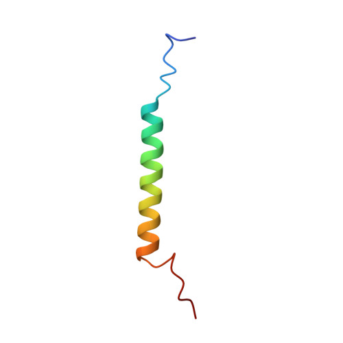
Bicelle-embedded integrin alpha(IIB) transmembrane segment
- PDB DOI: https://doi.org/10.2210/pdb2K1A/pdb
- Classification: CELL ADHESION
- Organism(s): Homo sapiens
- Expression System: Escherichia coli
- Mutation(s): No
- Membrane Protein: Yes OPMPDBTMMemProtMD
- Deposited: 2008-02-25 Released: 2008-04-15
- Deposition Author(s): Lau, T.-L., Dua, V., Ulmer, T.S.
Experimental Data Snapshot
- Method: SOLUTION NMR
- Conformers Calculated: 21
- Conformers Submitted: 21
- Selection Criteria: all calculated structures submitted
wwPDB Validation3D Report Full Report
Literature
- 👁 Image
Download Mendeley
Structure of the Integrin {alpha}IIb Transmembrane Segment.
Lau, T.L., Dua, V., Ulmer, T.S.(2008) J Biological Chem 283: 16162-16168
- PubMed: 18417472 Search on PubMedSearch on PubMed Central
- DOI: https://doi.org/10.1074/jbc.M801748200
- Primary Citation Related Structures:
2K1A - PubMed Abstract:
Integrin cell-adhesion receptors transduce signals bidirectionally across the plasma membrane via the single-pass transmembrane segments of each alpha and beta subunit. While the beta3 transmembrane segment consists of a linear 29-residue alpha-helix, the structure of the alphaIIb transmembrane segment reveals a linear 24-residue alpha-helix (Ile-966 -Lys-989) followed by a backbone reversal that packs Phe-992-Phe-993 against the transmembrane helix. The length of the alphaIIb transmembrane helix implies the absence of a significant transmembrane helix tilt in contrast to its partnering beta3 subunit. Sequence alignment shows Gly-991-Phe-993 to be fully conserved among all 18 human integrin alpha subunits, suggesting that their unusual structural motif is prototypical for integrin alpha subunits. The alphaIIb transmembrane structure demonstrates a level of complexity within the membrane that is beyond simple transmembrane helices and forms the structural basis for assessing the extent of structural and topological rearrangements upon alphaIIb-beta3 association, i.e. integrin transmembrane signaling.
- Department of Biochemistry and Molecular Biology and Zilkha Neurogenetic Institute, Keck School of Medicine, University of Southern California, Los Angeles, California 90033, USA.
Organizational Affiliation:
Explore in 3D: Structure | Sequence Annotations | Validation Report | Predict Membrane
Biological Assembly 1
Explore in 3D: Structure | Sequence Annotations | Validation Report | Predict Membrane
Global Symmetry: Asymmetric - C1
Global Stoichiometry: Monomer - A1
Find Similar Assemblies
Biological assembly 1 assigned by authors.
Macromolecule Content
- Total Structure Weight: 4.75 kDa
- Atom Count: 337
- Modeled Residue Count: 42
- Deposited Residue Count: 42
- Unique protein chains: 1
Macromolecules
Entity ID: 1 | |||||
|---|---|---|---|---|---|
| Molecule | Chains | Sequence Length | Organism | Details | Image |
| Integrin alpha-IIb | 42 | Homo sapiens | Mutation(s): 0 Gene Names: ITGA2B, GP2B, ITGAB Membrane Entity: Yes | 👁 Image | |
UniProt & NIH Common Fund Data Resources | |||||
PHAROS: P08514 GTEx: ENSG00000005961 | |||||
Entity Groups | |||||
| Sequence Clusters | 30% Identity50% Identity70% Identity90% Identity95% Identity100% Identity | ||||
| UniProt Group | P08514 | ||||
Sequence AnnotationsExpand | |||||
Reference Sequence | |||||
Experimental Data & Validation
Experimental Data
- Method: SOLUTION NMR
- Conformers Calculated: 21
- Conformers Submitted: 21
- Selection Criteria: all calculated structures submitted
Entry History
Deposition Data
- Released Date: 2008-04-15 Deposition Author(s): Lau, T.-L., Dua, V., Ulmer, T.S.
Revision History (Full details and data files)
- Version 1.0: 2008-04-15
Type: Initial release - Version 1.1: 2011-07-13
Changes: Version format compliance - Version 1.2: 2022-03-16
Changes: Data collection, Database references, Derived calculations - Version 1.3: 2024-05-29
Changes: Data collection