
- FASTA Sequence
- PDBx/mmCIF Format
- PDBx/mmCIF Format (gz)
- BinaryCIF Format (gz)
- Legacy PDB Format
- Legacy PDB Format (gz)
- PDBML/XML Format (gz)
- NMR Restraints (Text)
- NMR Restraints (Text - gz)
- NMR Restraints v2 (Text)
- NMR Restraints v2 (Text - gz)
- Validation Full (PDF - gz)
- Validation (XML - gz)
- Validation (CIF - gz)
- Biological Assembly 1 (CIF - gz)
- Biological Assembly 1 (PDB - gz)
2L1C | pdb_00002l1c
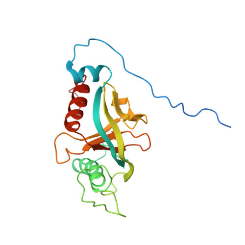
Shc-PTB:biphosphorylated integrin beta3 cytoplasmic tail complex (1:1)
- PDB DOI: https://doi.org/10.2210/pdb2L1C/pdb
- BMRB: 17080
- Classification: CELL ADHESION
- Organism(s): Homo sapiens
- Expression System: Escherichia coli
- Mutation(s): No
- Deposited: 2010-07-27 Released: 2010-08-18
- Deposition Author(s): Deshmukh, L., Gorbatyuk, V., Vinogradova, O.
Experimental Data Snapshot
- Method: SOLUTION NMR
- Conformers Calculated: 80
- Conformers Submitted: 15
- Selection Criteria: structures with the lowest energy
wwPDB Validation3D Report Full Report
Literature
- 👁 Image
Download Mendeley
Integrin {beta}3 phosphorylation dictates its complex with the Shc phosphotyrosine-binding (PTB) domain.
Deshmukh, L., Gorbatyuk, V., Vinogradova, O.(2010) J Biological Chem 285: 34875-34884
- PubMed: 20739287 Search on PubMedSearch on PubMed Central
- DOI: https://doi.org/10.1074/jbc.M110.159087
- Primary Citation Related Structures:
2L1C - PubMed Abstract:
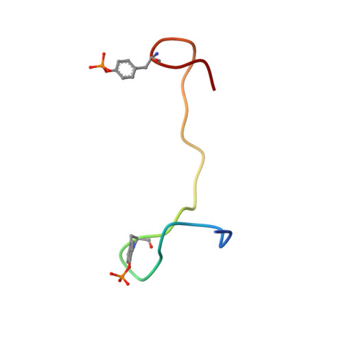
Adaptor protein Shc plays a key role in mitogen-activated protein kinase (MAPK) signaling pathway, which can be mediated through a number of different receptors including integrins. By specifically recognizing the tyrosine-phosphorylated integrin β(3), Shc has been shown to trigger integrin outside-in signaling, although the structural basis of this interaction remains nebulous. Here we present the detailed structural analysis of Shc phosphotyrosine-binding (PTB) domain in complex with the bi-phosphorylated β(3)integrin cytoplasmic tail (CT). We show that this complex is primarily defined by the phosphorylation state of the integrin C-terminal Tyr(759), which fits neatly into the classical PTB pocket of Shc. In addition, we have identified a novel binding interface which concurrently accommodates phosphorylated Tyr(747) of the highly conserved NPXY motif of β(3). The structure represents the first snapshot of an integrin cytoplasmic tail bound to a target for mediating the outside-in signaling. Detailed comparison with the known Shc PTB structure bound to a target TrkA peptide revealed some significant differences, which shed new light upon the PTB domain specificity.
- Department of Pharmaceutical Sciences, School of Pharmacy, University of Connecticut, Storrs, Connecticut 06269-3092, USA.
Organizational Affiliation:
Biological Assembly 1
Explore in 3D: Structure | Sequence Annotations | Validation Report
Global Symmetry: Asymmetric - C1
Global Stoichiometry: Hetero 2-mer - A1B1
Find Similar Assemblies
Biological assembly 1 assigned by authors.
Macromolecule Content
- Total Structure Weight: 26.45 kDa
- Atom Count: 1,697
- Modeled Residue Count: 218
- Deposited Residue Count: 238
- Unique protein chains: 2
Macromolecules
Entity ID: 1 | |||||
|---|---|---|---|---|---|
| Molecule | Chains | Sequence Length | Organism | Details | Image |
| SHC (Src homology 2 domain containing) transforming protein 1, isoform CRA_d | 211 | Homo sapiens | Mutation(s): 0 Gene Names: SHC1, hCG_1997126 | 👁 Image | |
UniProt & NIH Common Fund Data Resources | |||||
PHAROS: P29353 GTEx: ENSG00000160691 | |||||
Entity Groups | |||||
| Sequence Clusters | 30% Identity50% Identity70% Identity90% Identity95% Identity100% Identity | ||||
| UniProt Group | P29353 | ||||
Sequence AnnotationsExpand | |||||
Reference Sequence | |||||
Entity ID: 2 | |||||
|---|---|---|---|---|---|
| Molecule | Chains | Sequence Length | Organism | Details | Image |
| Integrin beta-3 | 27 | N/A | Mutation(s): 0 | 👁 Image | |
UniProt & NIH Common Fund Data Resources | |||||
PHAROS: P05106 GTEx: ENSG00000259207 | |||||
Entity Groups | |||||
| Sequence Clusters | 30% Identity50% Identity70% Identity90% Identity95% Identity100% Identity | ||||
| UniProt Group | P05106 | ||||
Sequence AnnotationsExpand | |||||
Reference Sequence | |||||
Small Molecules
| Modified Residues 1 Unique | |||||
|---|---|---|---|---|---|
| ID | Chains | Type | Formula | 2D Diagram | Parent |
| PTR Query on PTR | B | L-PEPTIDE LINKING | C9 H12 N O6 P | 👁 Image | TYR |
Experimental Data & Validation
Experimental Data
- Method: SOLUTION NMR
- Conformers Calculated: 80
- Conformers Submitted: 15
- Selection Criteria: structures with the lowest energy
Entry History
Deposition Data
- Released Date: 2010-08-18 Deposition Author(s): Deshmukh, L., Gorbatyuk, V., Vinogradova, O.
Revision History (Full details and data files)
- Version 1.0: 2010-08-18
Type: Initial release - Version 1.1: 2011-07-13
Changes: Version format compliance - Version 1.2: 2012-04-18
Changes: Database references - Version 1.3: 2024-10-09
Changes: Data collection, Database references, Derived calculations, Structure summary