
- FASTA Sequence
- PDBx/mmCIF Format
- PDBx/mmCIF Format (gz)
- BinaryCIF Format (gz)
- Legacy PDB Format
- Legacy PDB Format (gz)
- PDBML/XML Format (gz)
- Structure Factors (CIF)
- Structure Factors (CIF - gz)
- Validation Full (PDF - gz)
- Validation (XML - gz)
- Validation (CIF - gz)
- Validation 2fo-fc coefficients (CIF - gz)
- Validation fo-fc coefficients (CIF - gz)
- Biological Assembly 1 (CIF - gz)
- Biological Assembly 2 (CIF - gz)
- Biological Assembly 1 (PDB - gz)
- Biological Assembly 2 (PDB - gz)
2NZ1 | pdb_00002nz1
Viral Chemokine Binding Protein M3 From Murine Gammaherpesvirus68 In Complex With The CC-Chemokine CCL2/MCP-1
- PDB DOI: https://doi.org/10.2210/pdb2NZ1/pdb
- Classification: VIRAL PROTEIN/CYTOKINE
- Organism(s): Murid gammaherpesvirus 4, Homo sapiens
- Expression System: Spodoptera frugiperda, Escherichia coli
- Mutation(s): Yes
- Deposited: 2006-11-22 Released: 2007-12-25
- Deposition Author(s): Alexander-Brett, J.M., Fremont, D.H.
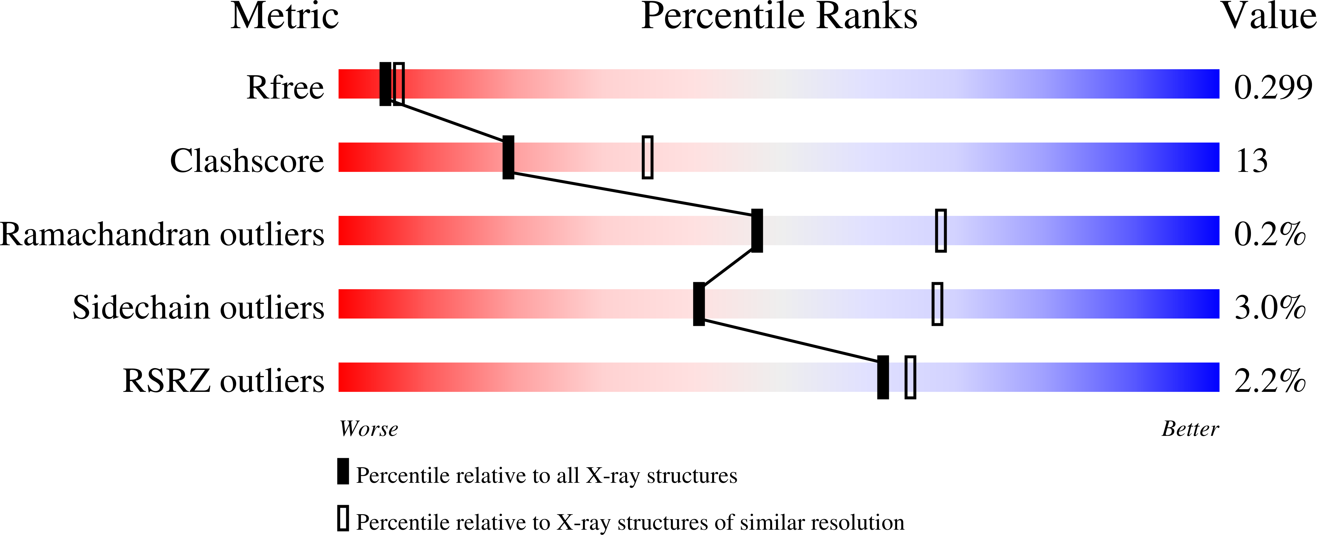
Experimental Data Snapshot
- Method: X-RAY DIFFRACTION
- Resolution: 2.50 Å
- R-Value Free: 0.300 (Depositor), 0.299 (DCC)
- R-Value Work: 0.231 (Depositor), 0.231 (DCC)
- R-Value Observed: 0.231 (Depositor)
Starting Model: experimental
View more details
wwPDB Validation3D Report Full Report
Literature
- 👁 Image
Download Mendeley
Dual GPCR and GAG mimicry by the M3 chemokine decoy receptor.
Alexander-Brett, J.M., Fremont, D.H.(2007) J Exp Medicine 204: 3157-3172
- PubMed: 18070938 Search on PubMedSearch on PubMed Central
- DOI: https://doi.org/10.1084/jem.20071677
- Primary Citation Related Structures:
2NYZ, 2NZ1 - PubMed Abstract:
Viruses have evolved a myriad of evasion strategies focused on undermining chemokine-mediated immune surveillance, exemplified by the mouse gamma-herpesvirus 68 M3 decoy receptor. Crystal structures of M3 in complex with C chemokine ligand 1/lymphotactin and CC chemokine ligand 2/monocyte chemoattractant protein 1 reveal that invariant chemokine features associated with G protein-coupled receptor binding are primarily recognized by the decoy C-terminal domain, whereas the N-terminal domain (NTD) reconfigures to engage divergent basic residue clusters on the surface of chemokines. Favorable electrostatic forces dramatically enhance the association kinetics of chemokine binding by M3, with a primary role ascribed to acidic NTD regions that effectively mimic glycosaminoglycan interactions. Thus, M3 employs two distinct mechanisms of chemical imitation to potently sequester chemokines, thereby inhibiting chemokine receptor binding events as well as the formation of chemotactic gradients necessary for directed leukocyte trafficking.
- Department of Pathology and Immunology, Washington University School of Medicine, St. Louis, MO 63110, USA.
Organizational Affiliation:
Explore in 3D: Structure | Sequence Annotations | Electron Density | Validation Report
Biological Assembly 1
Explore in 3D: Structure | Sequence Annotations | Electron Density | Validation Report
Global Symmetry: Cyclic - C2 (Explore in 3D)
Global Stoichiometry: Hetero 4-mer - A2B2
Find Similar Assemblies
Biological assembly 1 assigned by authors and generated by PISA,PQS (software)
Biological Assembly 2
Explore in 3D: Structure | Sequence Annotations | Electron Density | Validation Report
Global Symmetry: Cyclic - C2 (Explore in 3D)
Global Stoichiometry: Hetero 4-mer - A2B2
Find Similar Assemblies
Biological assembly 2 assigned by authors and generated by PQS (software)
Macromolecule Content
- Total Structure Weight: 151.52 kDa
- Atom Count: 10,672
- Modeled Residue Count: 1,305
- Deposited Residue Count: 1,374
- Unique protein chains: 2
Macromolecules
Entity ID: 1 | |||||
|---|---|---|---|---|---|
| Molecule | Chains | Sequence Length | Organism | Details | Image |
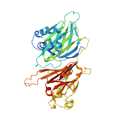
| Hypothetical protein GAMMAHV.M3 | A, B, E [auth X] | 382 | Murid gammaherpesvirus 4 | Mutation(s): 0 Gene Names: GAMMAHV.M3, M3 | 👁 Image |
UniProt | |||||
Entity Groups | |||||
| Sequence Clusters | 30% Identity50% Identity70% Identity90% Identity95% Identity100% Identity | ||||
| UniProt Group | O41925 | ||||
Sequence AnnotationsExpand | |||||
Reference Sequence | |||||
Entity ID: 2 | |||||
|---|---|---|---|---|---|
| Molecule | Chains | Sequence Length | Organism | Details | Image |
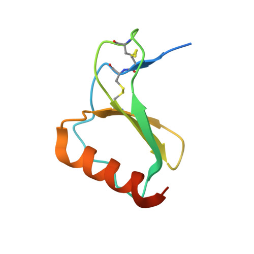
| Small inducible cytokine A2 | C [auth D], D [auth E], F [auth Y] | 76 | Homo sapiens | Mutation(s): 1 Gene Names: CCL2, MCP1, SCYA2 | 👁 Image |
UniProt & NIH Common Fund Data Resources | |||||
PHAROS: P13500 GTEx: ENSG00000108691 | |||||
Entity Groups | |||||
| Sequence Clusters | 30% Identity50% Identity70% Identity90% Identity95% Identity100% Identity | ||||
| UniProt Group | P13500 | ||||
Sequence AnnotationsExpand | |||||
Reference Sequence | |||||
Experimental Data & Validation
Experimental Data
- Method: X-RAY DIFFRACTION
- Resolution: 2.50 Å
- R-Value Free: 0.300 (Depositor), 0.299 (DCC)
- R-Value Work: 0.231 (Depositor), 0.231 (DCC)
- R-Value Observed: 0.231 (Depositor)
| Length ( Å ) | Angle ( ˚ ) |
|---|---|
| a = 99.24 | α = 90 |
| b = 99.24 | β = 90 |
| c = 243.46 | γ = 120 |
| Software Name | Purpose |
|---|---|
| CNS | refinement |
| HKL-2000 | data collection |
| HKL-2000 | data reduction |
| SCALEPACK | data scaling |
| AMoRE | phasing |
Entry History
Deposition Data
- Released Date: 2007-12-25 Deposition Author(s): Alexander-Brett, J.M., Fremont, D.H.
Revision History (Full details and data files)
- Version 1.0: 2007-12-25
Type: Initial release - Version 1.1: 2011-07-13
Changes: Derived calculations, Version format compliance - Version 1.2: 2021-10-20
Changes: Database references - Version 1.3: 2023-08-30
Changes: Data collection, Refinement description - Version 1.4: 2024-10-16
Changes: Structure summary