
- FASTA Sequence
- PDBx/mmCIF Format
- PDBx/mmCIF Format (gz)
- BinaryCIF Format (gz)
- Legacy PDB Format
- Legacy PDB Format (gz)
- PDBML/XML Format (gz)
- Structure Factors (CIF)
- Structure Factors (CIF - gz)
- Validation Full (PDF - gz)
- Validation (XML - gz)
- Validation (CIF - gz)
- Validation 2fo-fc coefficients (CIF - gz)
- Validation fo-fc coefficients (CIF - gz)
- Biological Assembly 1 (CIF - gz)
- Biological Assembly 1 (PDB - gz)
2OZ4 | pdb_00002oz4
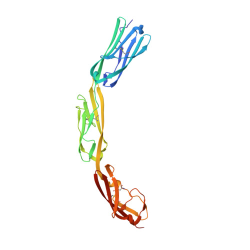
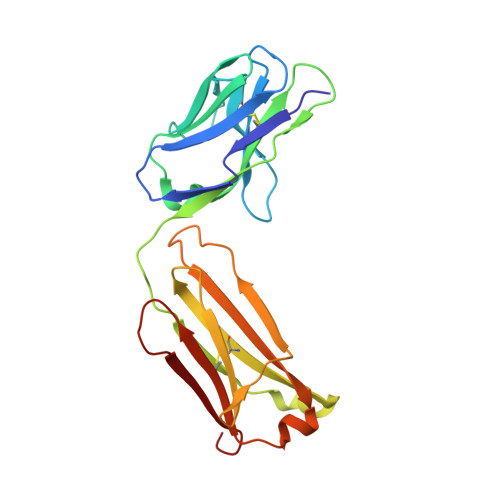
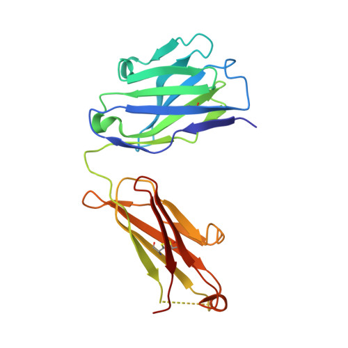
Structural Plasticity in IgSF Domain 4 of ICAM-1 Mediates Cell Surface Dimerization
- PDB DOI: https://doi.org/10.2210/pdb2OZ4/pdb
- Classification: CELL ADHESION
- Organism(s): Homo sapiens, Mus musculus
- Expression System: Cricetulus griseus
- Mutation(s): No
- Deposited: 2007-02-23 Released: 2007-10-16
- Deposition Author(s): Chen, X., Kim, T.D., Carman, C.V., Mi, L., Song, G., Springer, T.A.
Experimental Data Snapshot
- Method: X-RAY DIFFRACTION
- Resolution: 2.70 Å
- R-Value Free: 0.253 (Depositor), 0.250 (DCC)
- R-Value Work: 0.204 (Depositor), 0.200 (DCC)
- R-Value Observed: 0.209 (Depositor)
Starting Models: experimental
View more details
- 👁 Image
Download Mendeley
Structural plasticity in Ig superfamily domain 4 of ICAM-1 mediates cell surface dimerization.
Chen, X., Kim, T.D., Carman, C.V., Mi, L.Z., Song, G., Springer, T.A.(2007) Proc Natl Acad Sci U S A 104: 15358-15363
- PubMed: 17881562 Search on PubMedSearch on PubMed Central
- DOI: https://doi.org/10.1073/pnas.0707406104
- Primary Citation Related Structures:
2OZ4 - PubMed Abstract:
The Ig superfamily (IgSF) intercellular adhesion molecule-1 (ICAM-1) equilibrates between monomeric and dimeric forms on the cell surface, and dimerization enhances cell adhesion. A crystal structure of ICAM-1 IgSF domains (D) 3-5 revealed a unique dimerization interface in which D4s of two protomers fuse through edge beta-strands to form a single super beta-sandwich domain. Here, we describe a crystal structure at 2.7-A resolution of monomeric ICAM-1 D3-D5, stabilized by the monomer-specific Fab CA7. CA7 binds to D5 in a region that is buried in the dimeric interface and is distal from the dimerization site in D4. In monomeric ICAM-1 D3-D5, a 16-residue loop in D4 that is disordered in the dimeric structure could clearly be traced as a BC loop, a short C strand, and a CE meander with a cis-Pro followed by a solvent-exposed, flexible four-residue region. Deletions of 6 or 10 residues showed that the C-strand is essential for monomer stability, whereas a distinct six-residue deletion showed little contribution of the CE meander. Mutation of two inward-pointing Leu residues in edge beta-strand E to Lys increased monomer stability, confirming the hypothesis that inward-pointing charged side chains on edge beta-strands are an important design feature to prevent beta-supersheet formation. Overall, the studies reveal that monomer-dimer transition is associated with a surprisingly large, physiologically relevant, IgSF domain rearrangement.
- Immune Disease Institute, Department of Pathology, Harvard Medical School, Boston, MA 02115, USA.
Organizational Affiliation:
Explore in 3D: Structure | Sequence Annotations | Electron Density | Validation Report | Ligand Interaction (NAG)
Explore in 3D: Structure | Sequence Annotations | Electron Density | Validation Report | Ligand Interaction (NAG)
Global Symmetry: Asymmetric - C1
Global Stoichiometry: Hetero 3-mer - A1B1C1
Find Similar Assemblies
Biological assembly 1 assigned by authors and generated by PISA (software)
Macromolecule Content
- Total Structure Weight: 76.43 kDa
- Atom Count: 5,601
- Modeled Residue Count: 687
- Deposited Residue Count: 693
- Unique protein chains: 3
Entity ID: 1 | |||||
|---|---|---|---|---|---|
| Molecule | Chains | Sequence Length | Organism | Details | Image |
| Intercellular adhesion molecule 1 | 265 | Homo sapiens | Mutation(s): 0 Gene Names: ICAM1 | 👁 Image | |
UniProt & NIH Common Fund Data Resources | |||||
Find proteins for P05362 (Homo sapiens) Explore P05362 Go to UniProtKB: P05362 | |||||
PHAROS: P05362 GTEx: ENSG00000090339 | |||||
Entity Groups | |||||
| Sequence Clusters | 30% Identity50% Identity70% Identity90% Identity95% Identity100% Identity | ||||
| UniProt Group | P05362 | ||||
Glycosylation | |||||
| Glycosylation Sites: 3 | Go to GlyGen: P05362-1 | ||||
Sequence AnnotationsExpand | |||||
| |||||
Entity ID: 2 | |||||
|---|---|---|---|---|---|
| Molecule | Chains | Sequence Length | Organism | Details | Image |
| FAB FRAGMENT LIGHT CHAIN | B [auth L] | 214 | Mus musculus | Mutation(s): 0 | 👁 Image |
Entity Groups | |||||
| Sequence Clusters | 30% Identity50% Identity70% Identity90% Identity95% Identity100% Identity | ||||
Sequence AnnotationsExpand | |||||
| |||||
Entity ID: 3 | |||||
|---|---|---|---|---|---|
| Molecule | Chains | Sequence Length | Organism | Details | Image |
| FAB FRAGMENT, HEAVY CHAIN | C [auth H] | 214 | Mus musculus | Mutation(s): 0 | 👁 Image |
UniProt | |||||
Find proteins for P01757 (Mus musculus) Explore P01757 Go to UniProtKB: P01757 | |||||
Entity Groups | |||||
| Sequence Clusters | 30% Identity50% Identity70% Identity90% Identity95% Identity100% Identity | ||||
| UniProt Group | P01757 | ||||
Sequence AnnotationsExpand | |||||
| |||||
| Ligands 4 Unique | |||||
|---|---|---|---|---|---|
| ID | Chains | Name / Formula / InChI Key | 2D Diagram | 3D Interactions | |
| NAG Query on NAG Download Ideal Coordinates CCD File | E [auth A], F [auth A] | 2-acetamido-2-deoxy-beta-D-glucopyranose C8 H15 N O6 OVRNDRQMDRJTHS-FMDGEEDCSA-N | 👁 Image | ||
| TRS Query on TRS Download Ideal Coordinates CCD File | K [auth L], L | 2-AMINO-2-HYDROXYMETHYL-PROPANE-1,3-DIOL C4 H12 N O3 LENZDBCJOHFCAS-UHFFFAOYSA-O | 👁 Image | ||
| SO4 Query on SO4 Download Ideal Coordinates CCD File | G [auth A], H [auth L] | SULFATE ION O4 S QAOWNCQODCNURD-UHFFFAOYSA-L | 👁 Image | ||
| ZN Query on ZN Download Ideal Coordinates CCD File | I [auth L], J [auth L] | ZINC ION Zn PTFCDOFLOPIGGS-UHFFFAOYSA-N | 👁 Image | ||
Experimental Data
- Method: X-RAY DIFFRACTION
- Resolution: 2.70 Å
- R-Value Free: 0.253 (Depositor), 0.250 (DCC)
- R-Value Work: 0.204 (Depositor), 0.200 (DCC)
- R-Value Observed: 0.209 (Depositor)
| Length ( Å ) | Angle ( ˚ ) |
|---|---|
| a = 185.396 | α = 90 |
| b = 69.342 | β = 112.76 |
| c = 88.173 | γ = 90 |
| Software Name | Purpose |
|---|---|
| REFMAC | refinement |
| HKL-2000 | data collection |
| HKL-2000 | data reduction |
| HKL-2000 | data scaling |
| MOLREP | phasing |
Deposition Data
- Released Date: 2007-10-16 Deposition Author(s): Chen, X., Kim, T.D., Carman, C.V., Mi, L., Song, G., Springer, T.A.
Revision History (Full details and data files)
- Version 1.0: 2007-10-16
Type: Initial release - Version 1.1: 2011-07-13
Changes: Advisory, Version format compliance - Version 2.0: 2020-07-29
Type: Remediation
Reason: Carbohydrate remediation
Changes: Atomic model, Data collection, Derived calculations, Structure summary - Version 2.1: 2023-08-30
Changes: Data collection, Database references, Refinement description, Structure summary - Version 2.2: 2024-11-13
Changes: Structure summary