
- FASTA Sequence
- PDBx/mmCIF Format
- PDBx/mmCIF Format (gz)
- BinaryCIF Format (gz)
- Legacy PDB Format
- Legacy PDB Format (gz)
- PDBML/XML Format (gz)
- NMR Restraints (Text)
- NMR Restraints (Text - gz)
- NMR Restraints v2 (Text)
- NMR Restraints v2 (Text - gz)
- Validation Full (PDF - gz)
- Validation (XML - gz)
- Validation (CIF - gz)
- Biological Assembly 1 (CIF - gz)
- Biological Assembly 1 (PDB - gz)
2RNQ | pdb_00002rnq
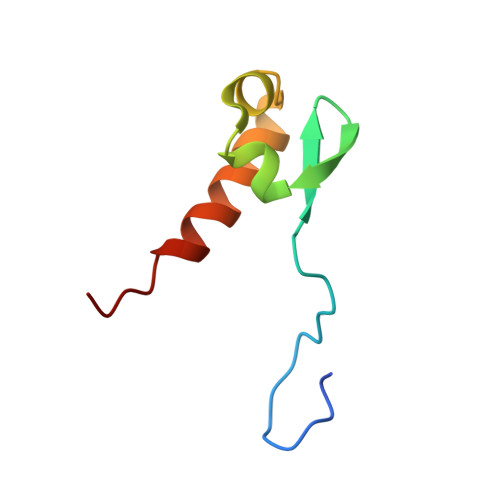
Solution structure of the C-terminal acidic domain of TFIIE alpha
- PDB DOI: https://doi.org/10.2210/pdb2RNQ/pdb
- Classification: TRANSCRIPTION
- Organism(s): Homo sapiens
- Expression System: Escherichia coli
- Mutation(s): No
- Deposited: 2008-01-31 Released: 2008-04-01
- Deposition Author(s): Okuda, M., Nishimura, Y.
Experimental Data Snapshot
- Method: SOLUTION NMR
- Conformers Calculated: 100
- Conformers Submitted: 20
- Selection Criteria: structures with the lowest energy
wwPDB Validation 3D Report Full Report
- 👁 Image
Download Mendeley
Structural insight into the TFIIE-TFIIH interaction: TFIIE and p53 share the binding region on TFIIH
Okuda, M., Tanaka, A., Satoh, M., Mizuta, S., Takazawa, M., Ohkuma, Y., Nishimura, Y.(2008) EMBO J 27: 1161-1171
- PubMed: 18354501 Search on PubMedSearch on PubMed Central
- DOI: https://doi.org/10.1038/emboj.2008.47
- Primary Citation Related Structures:
2RNQ, 2RNR - PubMed Abstract:
RNA polymerase II and general transcription factors (GTFs) assemble on a promoter to form a transcription preinitiation complex (PIC). Among the GTFs, TFIIE recruits TFIIH to complete the PIC formation and regulates enzymatic activities of TFIIH. However, the mode of binding between TFIIE and TFIIH is poorly understood. Here, we demonstrate the specific binding of the C-terminal acidic domain (AC-D) of the human TFIIEalpha subunit to the pleckstrin homology domain (PH-D) of the human TFIIH p62 subunit and describe the solution structures of the free and PH-D-bound forms of AC-D. Although the flexible N-terminal acidic tail from AC-D wraps around PH-D, the core domain of AC-D also interacts with PH-D. AC-D employs an entirely novel binding mode, which differs from the amphipathic helix method used by many transcriptional activators. So the binding surface between PH-D and AC-D is much broader than the specific binding surface between PH-D and the p53 acidic fragments. From our in vitro studies, we demonstrate that this interaction could be a switch to replace p53 with TFIIE on TFIIH in transcription.
- Laboratory of Structural Biology, Graduate School of Supramolecular Biology, Yokohama City University, Yokohama, Japan.
Organizational Affiliation:
Explore in 3D: Structure | Sequence Annotations | Validation Report
Global Symmetry: Asymmetric - C1
Global Stoichiometry: Monomer - A1
Find Similar Assemblies
Biological assembly 1 assigned by authors.
Macromolecule Content
- Total Structure Weight: 7.38 kDa
- Atom Count: 506
- Modeled Residue Count: 62
- Deposited Residue Count: 64
- Unique protein chains: 1
Entity ID: 1 | |||||
|---|---|---|---|---|---|
| Molecule | Chains | Sequence Length | Organism | Details | Image |
| Transcription initiation factor IIE subunit alpha | 64 | Homo sapiens | Mutation(s): 0 | 👁 Image | |
UniProt & NIH Common Fund Data Resources | |||||
Find proteins for P29083 (Homo sapiens) Explore P29083 Go to UniProtKB: P29083 | |||||
PHAROS: P29083 GTEx: ENSG00000153767 | |||||
Entity Groups | |||||
| Sequence Clusters | 30% Identity50% Identity70% Identity90% Identity95% Identity100% Identity | ||||
| UniProt Group | P29083 | ||||
Sequence AnnotationsExpand | |||||
| |||||
Experimental Data
- Method: SOLUTION NMR
- Conformers Calculated: 100
- Conformers Submitted: 20
- Selection Criteria: structures with the lowest energy
Deposition Data
- Released Date: 2008-04-01 Deposition Author(s): Okuda, M., Nishimura, Y.
Revision History (Full details and data files)
- Version 1.0: 2008-04-01
Type: Initial release - Version 1.1: 2011-07-13
Changes: Version format compliance - Version 1.2: 2022-03-16
Changes: Data collection, Database references, Derived calculations - Version 1.3: 2024-05-29
Changes: Data collection