
- FASTA Sequence
- PDBx/mmCIF Format
- PDBx/mmCIF Format (gz)
- BinaryCIF Format (gz)
- Legacy PDB Format
- Legacy PDB Format (gz)
- PDBML/XML Format (gz)
- Structure Factors (CIF)
- Structure Factors (CIF - gz)
- Validation Full (PDF - gz)
- Validation (XML - gz)
- Validation (CIF - gz)
- Validation 2fo-fc coefficients (CIF - gz)
- Validation fo-fc coefficients (CIF - gz)
- Biological Assembly 1 (CIF - gz)
- Biological Assembly 1 (PDB - gz)
2W0G | pdb_00002w0g
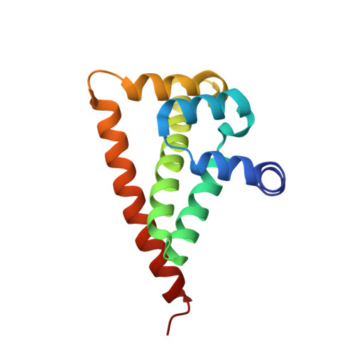
HSP90 CO-CHAPERONE CDC37
- PDB DOI: https://doi.org/10.2210/pdb2W0G/pdb
- Classification: CHAPERONE
- Organism(s): Homo sapiens
- Expression System: Escherichia coli BL21(DE3)
- Mutation(s): No
- Deposited: 2008-08-15 Released: 2008-12-09
- Deposition Author(s): Sreeramulu, S., Jonker, H.R.A., Schwalbe, H., Lancaster, C.R.D.
Experimental Data Snapshot
- Method: X-RAY DIFFRACTION
- Resolution: 1.88 Å
- R-Value Free: 0.246 (Depositor), 0.250 (DCC)
- R-Value Work: 0.212 (Depositor), 0.210 (DCC)
- R-Value Observed: 0.213 (Depositor)
Starting Model: experimental
View more details
wwPDB Validation 3D Report Full Report
- 👁 Image
Download Mendeley
The Human Cdc37.Hsp90 Complex Studied by Heteronuclear NMR Spectroscopy.
Sreeramulu, S., Jonker, H.R.A., Richter, C., Langer, T., Lancaster, C.R.D., Schwalbe, H.(2009) J Biological Chem 284: 3885
- PubMed: 19073599 Search on PubMed
- DOI: https://doi.org/10.1074/jbc.M806715200
- Primary Citation Related Structures:
2K5B, 2W0G - PubMed Abstract:
The cell division cycle protein 37 (Cdc37) and the 90-kDa heat shock protein (Hsp90) are molecular chaperones, which are crucial elements in the protein signaling pathway. The largest class of client proteins for Cdc37 and Hsp90 are protein kinases. The catalytic domains of these kinases are stabilized by Cdc37, and their proper folding and functioning is dependent on Hsp90. Here, we present the x-ray crystal structure of the 16-kDa middle domain of human Cdc37 at 1.88 angstroms resolution and the structure of this domain in complex with the 23-kDa N-terminal domain of human Hsp90 based on heteronuclear solution state NMR data and docking. Our results demonstrate that the middle domain of Cdc37 exists as a monomer. NMR and mutagenesis experiments reveal Leu-205 in Cdc37 as a key residue enabling complex formation. These findings can be very useful in the development of small molecule inhibitors against cancer.
- Institute for Organic Chemistry and Chemical Biology, Center for Biomolecular Magnetic Resonance, Johann Wolfgang Goethe-University, Max-von-Laue-Strasse 7, Frankfurt am Main D-60438, Germany.
Organizational Affiliation:
Explore in 3D: Structure | Sequence Annotations | Electron Density | Validation Report
Explore in 3D: Structure | Sequence Annotations | Electron Density | Validation Report
Global Symmetry: Asymmetric - C1
Global Stoichiometry: Monomer - A1
Find Similar Assemblies
Biological assembly 1 assigned by authors and generated by PQS (software)
Macromolecule Content
- Total Structure Weight: 15.52 kDa
- Atom Count: 1,180
- Modeled Residue Count: 129
- Deposited Residue Count: 129
- Unique protein chains: 1
Entity ID: 1 | |||||
|---|---|---|---|---|---|
| Molecule | Chains | Sequence Length | Organism | Details | Image |
| HSP90 CO-CHAPERONE CDC37 | 129 | Homo sapiens | Mutation(s): 0 | 👁 Image | |
UniProt & NIH Common Fund Data Resources | |||||
Find proteins for Q16543 (Homo sapiens) Explore Q16543 Go to UniProtKB: Q16543 | |||||
PHAROS: Q16543 GTEx: ENSG00000105401 | |||||
Entity Groups | |||||
| Sequence Clusters | 30% Identity50% Identity70% Identity90% Identity95% Identity100% Identity | ||||
| UniProt Group | Q16543 | ||||
Sequence AnnotationsExpand | |||||
| |||||
Experimental Data
- Method: X-RAY DIFFRACTION
- Resolution: 1.88 Å
- R-Value Free: 0.246 (Depositor), 0.250 (DCC)
- R-Value Work: 0.212 (Depositor), 0.210 (DCC)
- R-Value Observed: 0.213 (Depositor)
| Length ( Å ) | Angle ( ˚ ) |
|---|---|
| a = 48.67 | α = 90 |
| b = 48.67 | β = 90 |
| c = 104.257 | γ = 120 |
| Software Name | Purpose |
|---|---|
| REFMAC | refinement |
| DENZO | data reduction |
| SCALEPACK | data scaling |
| AMoRE | phasing |
Deposition Data
- Released Date: 2008-12-09 Deposition Author(s): Sreeramulu, S., Jonker, H.R.A., Schwalbe, H., Lancaster, C.R.D.
Revision History (Full details and data files)
- Version 1.0: 2008-12-09
Type: Initial release - Version 1.1: 2011-05-08
Changes: Version format compliance - Version 1.2: 2011-07-13
Changes: Version format compliance - Version 1.3: 2017-07-05
Changes: Data collection - Version 1.4: 2023-12-13
Changes: Advisory, Data collection, Database references, Other, Refinement description