
- FASTA Sequence
- PDBx/mmCIF Format
- PDBx/mmCIF Format (gz)
- BinaryCIF Format (gz)
- Legacy PDB Format
- Legacy PDB Format (gz)
- PDBML/XML Format (gz)
- Structure Factors (CIF)
- Structure Factors (CIF - gz)
- Validation Full (PDF - gz)
- Validation (XML - gz)
- Validation (CIF - gz)
- Validation 2fo-fc coefficients (CIF - gz)
- Validation fo-fc coefficients (CIF - gz)
- Biological Assembly 1 (CIF - gz)
- Biological Assembly 1 (PDB - gz)
2WWX | pdb_00002wwx
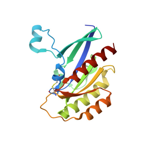
Crystal structure of the SidM/DrrA(GEF/GDF domain)-Rab1(GTPase domain) complex
- PDB DOI: https://doi.org/10.2210/pdb2WWX/pdb
- Classification: PROTEIN TRANSPORT
- Organism(s): Homo sapiens, Legionella pneumophila
- Expression System: Escherichia coli BL21(DE3)
- Mutation(s): Yes
- Deposited: 2009-10-30 Released: 2009-12-08
- Deposition Author(s): Suh, H.Y., Lee, D.W., Woo, J.S., Oh, B.H.
Experimental Data Snapshot
- Method: X-RAY DIFFRACTION
- Resolution: 1.50 Å
- R-Value Free: 0.231 (Depositor)
- R-Value Work: 0.212 (Depositor), 0.211 (DCC)
- R-Value Observed: 0.213 (Depositor)
wwPDB Validation 3D Report Full Report
- 👁 Image
Download Mendeley
Structural Insights Into the Dual Nucleotide Exchange and Gdi Displacement Activity of Sidm/Drra
Suh, H.Y., Lee, D.W., Lee, K.H., Ku, B., Choi, S.J., Woo, J.S., Kim, Y.G., Oh, B.H.(2010) EMBO J 29: 496
- PubMed: 19942850 Search on PubMedSearch on PubMed Central
- DOI: https://doi.org/10.1038/emboj.2009.347
- Primary Citation Related Structures:
2WWX - PubMed Abstract:
GDP-bound prenylated Rabs, sequestered by GDI (GDP dissociation inhibitor) in the cytosol, are delivered to destined sub-cellular compartment and subsequently activated by GEFs (guanine nucleotide exchange factors) catalysing GDP-to-GTP exchange. The dissociation of GDI from Rabs is believed to require a GDF (GDI displacement factor). Only two RabGDFs, human PRA-1 and Legionella pneumophila SidM/DrrA, have been identified so far and the molecular mechanism of GDF is elusive. Here, we present the structure of a SidM/DrrA fragment possessing dual GEF and GDF activity in complex with Rab1. SidM/DrrA reconfigures the Switch regions of the GTPase domain of Rab1, as eukaryotic GEFs do toward cognate Rabs. Structure-based mutational analyses show that the surface of SidM/DrrA, catalysing nucleotide exchange, is involved in GDI1 displacement from prenylated Rab1:GDP. In comparison with an eukaryotic GEF TRAPP I, this bacterial GEF/GDF exhibits high binding affinity for Rab1 with GDP retained at the active site, which appears as the key feature for the GDF activity of the protein.
- Department of Life Sciences and Center for Biomolecular Recognition, Pohang University of Science and Technology, Pohang, Kyungbuk, Korea.
Organizational Affiliation:
Explore in 3D: Structure | Sequence Annotations | Electron Density | Validation Report
Explore in 3D: Structure | Sequence Annotations | Electron Density | Validation Report
Global Symmetry: Asymmetric - C1
Global Stoichiometry: Hetero 2-mer - A1B1
Find Similar Assemblies
Biological assembly 1 generated by PISA (software)
Macromolecule Content
- Total Structure Weight: 44.17 kDa
- Atom Count: 3,038
- Modeled Residue Count: 370
- Deposited Residue Count: 392
- Unique protein chains: 2
Entity ID: 1 | |||||
|---|---|---|---|---|---|
| Molecule | Chains | Sequence Length | Organism | Details | Image |
| RAS-RELATED PROTEIN RAB-1 | 175 | Homo sapiens | Mutation(s): 1 EC: 3.6.5.2 | 👁 Image | |
UniProt & NIH Common Fund Data Resources | |||||
Find proteins for P62820 (Homo sapiens) Explore P62820 Go to UniProtKB: P62820 | |||||
PHAROS: P62820 GTEx: ENSG00000138069 | |||||
Entity Groups | |||||
| Sequence Clusters | 30% Identity50% Identity70% Identity90% Identity95% Identity100% Identity | ||||
| UniProt Group | P62820 | ||||
Sequence AnnotationsExpand | |||||
| |||||
Entity ID: 2 | |||||
|---|---|---|---|---|---|
| Molecule | Chains | Sequence Length | Organism | Details | Image |
| DRRA | 217 | Legionella pneumophila | Mutation(s): 0 EC: 2.7.7.108 | 👁 Image | |
UniProt | |||||
Find proteins for Q5ZSQ3 (Legionella pneumophila subsp. pneumophila (strain Philadelphia 1 / ATCC 33152 / DSM 7513)) Explore Q5ZSQ3 Go to UniProtKB: Q5ZSQ3 | |||||
Entity Groups | |||||
| Sequence Clusters | 30% Identity50% Identity70% Identity90% Identity95% Identity100% Identity | ||||
| UniProt Group | Q5ZSQ3 | ||||
Sequence AnnotationsExpand | |||||
| |||||
Experimental Data
- Method: X-RAY DIFFRACTION
- Resolution: 1.50 Å
- R-Value Free: 0.231 (Depositor)
- R-Value Work: 0.212 (Depositor), 0.211 (DCC)
- R-Value Observed: 0.213 (Depositor)
| Length ( Å ) | Angle ( ˚ ) |
|---|---|
| a = 68.88 | α = 90 |
| b = 73.803 | β = 90 |
| c = 73.665 | γ = 90 |
| Software Name | Purpose |
|---|---|
| REFMAC | refinement |
| HKL-2000 | data reduction |
| SCALEPACK | data scaling |
Deposition Data
Revision History (Full details and data files)
- Version 1.0: 2009-12-08
Type: Initial release - Version 1.1: 2015-04-01
Changes: Data collection, Version format compliance - Version 1.2: 2024-11-06
Changes: Data collection, Database references, Other, Structure summary