
- FASTA Sequence
- PDBx/mmCIF Format
- PDBx/mmCIF Format (gz)
- BinaryCIF Format (gz)
- Legacy PDB Format
- Legacy PDB Format (gz)
- PDBML/XML Format (gz)
- Structure Factors (CIF)
- Structure Factors (CIF - gz)
- Validation Full (PDF - gz)
- Validation (XML - gz)
- Validation (CIF - gz)
- Validation 2fo-fc coefficients (CIF - gz)
- Validation fo-fc coefficients (CIF - gz)
- Biological Assembly 1 (CIF - gz)
- Biological Assembly 1 (PDB - gz)
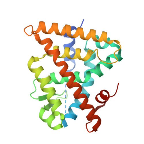
3E94 | pdb_00003e94
Crystal structure of RXRalpha ligand binding domain in complex with tributyltin and a coactivator fragment
- PDB DOI: https://doi.org/10.2210/pdb3E94/pdb
- Classification: TRANSCRIPTION
- Organism(s): Homo sapiens
- Expression System: Escherichia coli
- Mutation(s): No
- Deposited: 2008-08-21 Released: 2009-03-10
- Deposition Author(s): Bourguet, W., Le Maire, A.
Experimental Data Snapshot
- Method: X-RAY DIFFRACTION
- Resolution: 1.90 Å
- R-Value Free: 0.225 (Depositor), 0.230 (DCC)
- R-Value Work: 0.190 (Depositor), 0.190 (DCC)
- R-Value Observed: 0.192 (Depositor)
Starting Model: experimental
View more details
- 👁 Image
Download Mendeley
Activation of RXR-PPAR heterodimers by organotin environmental endocrine disruptors
le Maire, A., Grimaldi, M., Roecklin, D., Dagnino, S., Vivat-Hannah, V., Balaguer, P., Bourguet, W.(2009) EMBO Rep 10: 367-373
- PubMed: 19270714 Search on PubMedSearch on PubMed Central
- DOI: https://doi.org/10.1038/embor.2009.8
- Primary Citation Related Structures:
3E94 - PubMed Abstract:
The nuclear receptor retinoid X receptor-alpha (RXR-alpha)-peroxisome proliferator-activated receptor-gamma (PPAR-gamma) heterodimer was recently reported to have a crucial function in mediating the deleterious effects of organotin compounds, which are ubiquitous environmental contaminants. However, because organotins are unrelated to known RXR-alpha and PPAR-gamma ligands, the mechanism by which these compounds bind to and activate the RXR-alpha-PPAR-gamma heterodimer at nanomolar concentrations has remained elusive. Here, we show that tributyltin (TBT) activates all three RXR-PPAR-alpha, -gamma, -delta heterodimers, primarily through its interaction with RXR. In addition, the 1.9 A resolution structure of the RXR-alpha ligand-binding domain in complex with TBT shows a covalent bond between the tin atom and residue Cys 432 of helix H11. This interaction largely accounts for the high binding affinity of TBT, which only partly occupies the RXR-alpha ligand-binding pocket. Our data allow an understanding of the binding and activation properties of the various organotins and suggest a mechanism by which these tin compounds could affect other nuclear receptor signalling pathways.
- INSERM, U554, Universités Montpellier 1 & 2, 29 rue de Navacelles, 34090 Montpellier, France.
Organizational Affiliation:
Explore in 3D: Structure | Sequence Annotations | Electron Density | Validation Report | Ligand Interaction (TBY)
Explore in 3D: Structure | Sequence Annotations | Electron Density | Validation Report | Ligand Interaction (TBY)
Global Symmetry: Cyclic - C2 (Explore in 3D)
Global Stoichiometry: Hetero 4-mer - A2B2
Pseudo Symmetry: Asymmetric - C1
Pseudo Stoichiometry: Hetero 4-mer - A2B1C1
Find Similar Assemblies
Biological assembly 1 assigned by authors.
Macromolecule Content
- Total Structure Weight: 29.2 kDa
- Atom Count: 1,956
- Modeled Residue Count: 221
- Deposited Residue Count: 257
- Unique protein chains: 2
Entity ID: 1 | |||||
|---|---|---|---|---|---|
| Molecule | Chains | Sequence Length | Organism | Details | Image |
| Retinoic acid receptor RXR-alpha | 244 | Homo sapiens | Mutation(s): 0 Gene Names: RXRA, NR2B1 | 👁 Image | |
UniProt & NIH Common Fund Data Resources | |||||
Find proteins for P19793 (Homo sapiens) Explore P19793 Go to UniProtKB: P19793 | |||||
PHAROS: P19793 GTEx: ENSG00000186350 | |||||
Entity Groups | |||||
| Sequence Clusters | 30% Identity50% Identity70% Identity90% Identity95% Identity100% Identity | ||||
| UniProt Group | P19793 | ||||
Sequence AnnotationsExpand | |||||
| |||||
Find similar proteins by: Sequence | 3D Structure
Entity ID: 2 | |||||
|---|---|---|---|---|---|
| Molecule | Chains | Sequence Length | Organism | Details | Image |
| Nuclear receptor coactivator 2 peptide | 13 | N/A | Mutation(s): 0 | 👁 Image | |
UniProt & NIH Common Fund Data Resources | |||||
Find proteins for Q15596 (Homo sapiens) Explore Q15596 Go to UniProtKB: Q15596 | |||||
PHAROS: Q15596 GTEx: ENSG00000140396 | |||||
Entity Groups | |||||
| Sequence Clusters | 30% Identity50% Identity70% Identity90% Identity95% Identity100% Identity | ||||
| UniProt Group | Q15596 | ||||
Sequence AnnotationsExpand | |||||
| |||||
| Ligands 2 Unique | |||||
|---|---|---|---|---|---|
| ID | Chains | Name / Formula / InChI Key | 2D Diagram | 3D Interactions | |
| TBY Query on TBY Download Ideal Coordinates CCD File | C [auth A] | tributylstannanyl C12 H27 Sn DBGVGMSCBYYSLD-UHFFFAOYSA-N | 👁 Image | ||
| ACT Query on ACT Download Ideal Coordinates CCD File | D [auth A] | ACETATE ION C2 H3 O2 QTBSBXVTEAMEQO-UHFFFAOYSA-M | 👁 Image | ||
Experimental Data
- Method: X-RAY DIFFRACTION
- Resolution: 1.90 Å
- R-Value Free: 0.225 (Depositor), 0.230 (DCC)
- R-Value Work: 0.190 (Depositor), 0.190 (DCC)
- R-Value Observed: 0.192 (Depositor)
| Length ( Å ) | Angle ( ˚ ) |
|---|---|
| a = 64.028 | α = 90 |
| b = 64.028 | β = 90 |
| c = 111.882 | γ = 90 |
| Software Name | Purpose |
|---|---|
| MOSFLM | data reduction |
| SCALA | data scaling |
| REFMAC | refinement |
| PDB_EXTRACT | data extraction |
| ADSC | data collection |
| REFMAC | phasing |
Deposition Data
- Released Date: 2009-03-10 Deposition Author(s): Bourguet, W., Le Maire, A.
Revision History (Full details and data files)
- Version 1.0: 2009-03-10
Type: Initial release - Version 1.1: 2011-07-13
Changes: Version format compliance - Version 1.2: 2022-08-17
Changes: Database references, Derived calculations - Version 1.3: 2023-08-30
Changes: Data collection, Refinement description