
- FASTA Sequence
- PDBx/mmCIF Format
- PDBx/mmCIF Format (gz)
- BinaryCIF Format (gz)
- Legacy PDB Format
- Legacy PDB Format (gz)
- PDBML/XML Format (gz)
- Structure Factors (CIF)
- Structure Factors (CIF - gz)
- Validation Full (PDF - gz)
- Validation (XML - gz)
- Validation (CIF - gz)
- Validation 2fo-fc coefficients (CIF - gz)
- Validation fo-fc coefficients (CIF - gz)
- Biological Assembly 1 (CIF - gz)
- Biological Assembly 2 (CIF - gz)
- Biological Assembly 3 (CIF - gz)
- Biological Assembly 1 (PDB - gz)
- Biological Assembly 2 (PDB - gz)
- Biological Assembly 3 (PDB - gz)
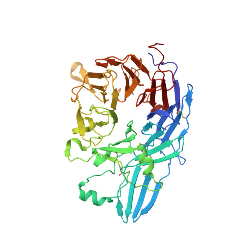
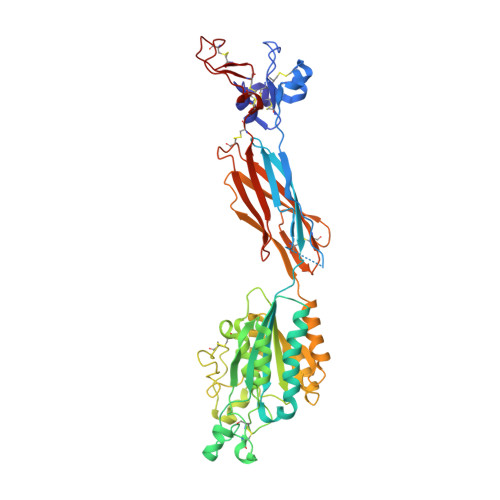
3FCU | pdb_00003fcu
Structure of headpiece of integrin aIIBb3 in open conformation
- PDB DOI: https://doi.org/10.2210/pdb3FCU/pdb
- Classification: Cell Adhesion/BLOOD CLOTTING
- Organism(s): Homo sapiens
- Expression System: Cricetulus griseus
- Mutation(s): No
- Deposited: 2008-11-22 Released: 2009-01-20
- Deposition Author(s): Zhu, J., Luo, B.-H., Xiao, T., Zhang, C., Nishida, N., Springer, T.A.
Experimental Data Snapshot
- Method: X-RAY DIFFRACTION
- Resolution: 2.90 Å
- R-Value Free: 0.197 (Depositor), 0.197 (DCC)
- R-Value Work: 0.174 (Depositor), 0.177 (DCC)
- R-Value Observed: 0.174 (Depositor)
Starting Model: experimental
View more details
Literature
- 👁 Image
Download Mendeley
Structure of a complete integrin ectodomain in a physiologic resting state and activation and deactivation by applied forces.
Zhu, J., Luo, B.H., Xiao, T., Zhang, C., Nishida, N., Springer, T.A.(2008) Mol Cell 32: 849-861
- PubMed: 19111664 Search on PubMedSearch on PubMed Central
- DOI: https://doi.org/10.1016/j.molcel.2008.11.018
- Primary Citation Related Structures:
3FCS, 3FCU - PubMed Abstract:
The complete ectodomain of integrin alpha(IIb)beta(3) reveals a bent, closed, low-affinity conformation, the beta knee, and a mechanism for linking cytoskeleton attachment to high affinity for ligand. Ca and Mg ions in the recognition site, including the synergistic metal ion binding site (SyMBS), are loaded prior to ligand binding. Electrophilicity of the ligand-binding Mg ion is increased in the open conformation. The beta(3) knee passes between the beta(3)-PSI and alpha(IIb)-knob to bury the lower beta leg in a cleft, from which it is released for extension. Different integrin molecules in crystals and EM reveal breathing that appears on pathway to extension. Tensile force applied to the extended ligand-receptor complex stabilizes the closed, low-affinity conformation. By contrast, an additional lateral force applied to the beta subunit to mimic attachment to moving actin filaments stabilizes the open, high-affinity conformation. This mechanism propagates allostery over long distances and couples cytoskeleton attachment of integrins to their high-affinity state.
- The Immune Disease Institute and Department of Pathology, Harvard Medical School, 200 Longwood Avenue, Boston, MA 02115, USA.
Organizational Affiliation:
Explore in 3D: Structure | Sequence Annotations | Electron Density | Validation Report | Ligand Interaction (NAG)
Biological Assembly 1
Explore in 3D: Structure | Sequence Annotations | Electron Density | Validation Report | Ligand Interaction (NAG)
Global Symmetry: Asymmetric - C1
Global Stoichiometry: Hetero 2-mer - A1B1
Find Similar Assemblies
Biological assembly 1 assigned by authors and generated by PISA (software)
Biological Assembly 2
Explore in 3D: Structure | Sequence Annotations | Electron Density | Validation Report | Ligand Interaction (NAG)
Global Symmetry: Asymmetric - C1
Global Stoichiometry: Hetero 2-mer - A1B1
Find Similar Assemblies
Biological assembly 2 assigned by authors and generated by PISA (software)
Biological Assembly 3
Explore in 3D: Structure | Sequence Annotations | Electron Density | Validation Report | Ligand Interaction (NAG)
Global Symmetry: Asymmetric - C1
Global Stoichiometry: Hetero 2-mer - A1B1
Find Similar Assemblies
Biological assembly 3 assigned by authors and generated by PISA (software)
Macromolecule Content
- Total Structure Weight: 307.08 kDa
- Atom Count: 21,657
- Modeled Residue Count: 2,725
- Deposited Residue Count: 2,754
- Unique protein chains: 2
Macromolecules
Entity ID: 1 | |||||
|---|---|---|---|---|---|
| Molecule | Chains | Sequence Length | Organism | Details | Image |
| Integrin, alpha 2b | 457 | Homo sapiens | Mutation(s): 0 Gene Names: ITGA2B | 👁 Image | |
UniProt & NIH Common Fund Data Resources | |||||
PHAROS: P08514 GTEx: ENSG00000005961 | |||||
Entity Groups | |||||
| Sequence Clusters | 30% Identity50% Identity70% Identity90% Identity95% Identity100% Identity | ||||
| UniProt Group | P08514 | ||||
Glycosylation | |||||
| Glycosylation Sites: 1 | Go to GlyGen: P08514-1 | ||||
Sequence AnnotationsExpand | |||||
Reference Sequence | |||||
Entity ID: 2 | |||||
|---|---|---|---|---|---|
| Molecule | Chains | Sequence Length | Organism | Details | Image |
| Integrin beta-3 | 461 | Homo sapiens | Mutation(s): 0 Gene Names: ITGB3, GP3A | 👁 Image | |
UniProt & NIH Common Fund Data Resources | |||||
PHAROS: P05106 GTEx: ENSG00000259207 | |||||
Entity Groups | |||||
| Sequence Clusters | 30% Identity50% Identity70% Identity90% Identity95% Identity100% Identity | ||||
| UniProt Group | P05106 | ||||
Glycosylation | |||||
| Glycosylation Sites: 3 | Go to GlyGen: P05106-1 | ||||
Sequence AnnotationsExpand | |||||
Reference Sequence | |||||
Oligosaccharides
HelpEntity ID: 3 | |||||
|---|---|---|---|---|---|
| Molecule | Chains | Length | 2D Diagram | Glycosylation | D Interactions |
| alpha-D-mannopyranose-(1-3)-[alpha-D-mannopyranose-(1-6)]alpha-D-mannopyranose-(1-4)-2-acetamido-2-deoxy-beta-D-glucopyranose-(1-4)-2-acetamido-2-deoxy-beta-D-glucopyranose | G, I | 5 | 👁 Image | N-Glycosylation | |
Glycosylation Resources | |||||
GlyTouCan: G21381MC GlyCosmos: G21381MC GlyGen: G21381MC | |||||
Entity ID: 4 | |||||
|---|---|---|---|---|---|
| Molecule | Chains | Length | 2D Diagram | Glycosylation | D Interactions |
| alpha-D-mannopyranose-(1-3)-alpha-D-mannopyranose-(1-4)-2-acetamido-2-deoxy-beta-D-glucopyranose-(1-4)-2-acetamido-2-deoxy-beta-D-glucopyranose | H | 4 | 👁 Image | N-Glycosylation | |
Glycosylation Resources | |||||
GlyTouCan: G48068RF GlyCosmos: G48068RF GlyGen: G48068RF | |||||
Small Molecules
Experimental Data & Validation
Experimental Data
- Method: X-RAY DIFFRACTION
- Resolution: 2.90 Å
- R-Value Free: 0.197 (Depositor), 0.197 (DCC)
- R-Value Work: 0.174 (Depositor), 0.177 (DCC)
- R-Value Observed: 0.174 (Depositor)
| Length ( Å ) | Angle ( ˚ ) |
|---|---|
| a = 332.093 | α = 90 |
| b = 332.093 | β = 90 |
| c = 88.288 | γ = 120 |
| Software Name | Purpose |
|---|---|
| ADSC | data collection |
| AMoRE | phasing |
| REFMAC | refinement |
| HKL-2000 | data reduction |
| SCALEPACK | data scaling |
Entry History
Deposition Data
- Released Date: 2009-01-20 Deposition Author(s): Zhu, J., Luo, B.-H., Xiao, T., Zhang, C., Nishida, N., Springer, T.A.
Revision History (Full details and data files)
- Version 1.0: 2009-01-20
Type: Initial release - Version 1.1: 2011-07-13
Changes: Advisory, Version format compliance - Version 2.0: 2020-07-29
Type: Remediation
Reason: Carbohydrate remediation
Changes: Advisory, Atomic model, Data collection, Derived calculations, Structure summary - Version 2.1: 2023-09-06
Changes: Data collection, Database references, Refinement description, Structure summary - Version 2.2: 2024-11-27
Changes: Structure summary