
- FASTA Sequence
- PDBx/mmCIF Format
- PDBx/mmCIF Format (gz)
- BinaryCIF Format (gz)
- Legacy PDB Format
- Legacy PDB Format (gz)
- PDBML/XML Format (gz)
- Structure Factors (CIF)
- Structure Factors (CIF - gz)
- Validation Full (PDF - gz)
- Validation (XML - gz)
- Validation (CIF - gz)
- Validation 2fo-fc coefficients (CIF - gz)
- Validation fo-fc coefficients (CIF - gz)
- Biological Assembly 1 (CIF - gz)
- Biological Assembly 2 (CIF - gz)
- Biological Assembly 3 (CIF - gz)
- Biological Assembly 4 (CIF - gz)
- Biological Assembly 1 (PDB - gz)
- Biological Assembly 2 (PDB - gz)
- Biological Assembly 3 (PDB - gz)
- Biological Assembly 4 (PDB - gz)
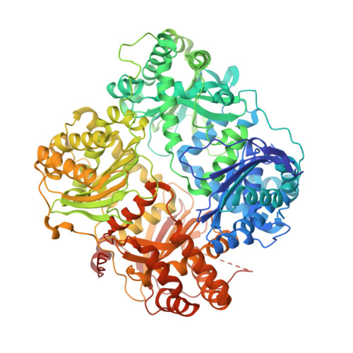
3H44 | pdb_00003h44
Crystal Structure of Insulin Degrading Enzyme in Complex with macrophage inflammatory protein 1 alpha
- PDB DOI: https://doi.org/10.2210/pdb3H44/pdb
- Classification: Hydrolase/Cytokine
- Organism(s): Homo sapiens
- Expression System: Escherichia coli
- Mutation(s): Yes
- Deposited: 2009-04-17 Released: 2010-04-14
- Deposition Author(s): Ren, M., Guo, Q., Tang, W.J.
Experimental Data Snapshot
- Method: X-RAY DIFFRACTION
- Resolution: 3.00 Å
- R-Value Free: 0.237 (Depositor), 0.240 (DCC)
- R-Value Work: 0.183 (Depositor), 0.190 (DCC)
- R-Value Observed: 0.185 (Depositor)
Starting Model: experimental
View more details
wwPDB Validation 3D Report Full Report
- 👁 Image
Download Mendeley
Macrophage Inflammatory Protein-1 Is A Novel High Affinity Substrate For Human Insulin Degrading Enzyme
Ren, M., Guo, Q., Tang, W.J.To be published.
Explore in 3D: Structure | Sequence Annotations | Electron Density | Validation Report | Ligand Interaction (DIO)
Explore in 3D: Structure | Sequence Annotations | Electron Density | Validation Report | Ligand Interaction (DIO)
Global Symmetry: Cyclic - C2 (Explore in 3D)
Global Stoichiometry: Hetero 4-mer - A2B2
Pseudo Symmetry: Asymmetric - C1
Pseudo Stoichiometry: Hetero 4-mer - A2B1C1
Find Similar Assemblies
Biological assembly 1 generated by PISA (software)
Explore in 3D: Structure | Sequence Annotations | Electron Density | Validation Report | Ligand Interaction (DIO)
Global Symmetry: Asymmetric - C1
Global Stoichiometry: Hetero 4-mer - A2B2
Pseudo Symmetry: Asymmetric - C1
Pseudo Stoichiometry: Hetero 4-mer - A2B1C1
Find Similar Assemblies
Biological assembly 2 assigned by authors and generated by PISA (software)
Explore in 3D: Structure | Sequence Annotations | Electron Density | Validation Report | Ligand Interaction (DIO)
Global Symmetry: Asymmetric - C1
Global Stoichiometry: Hetero 2-mer - A1B1
Find Similar Assemblies
Biological assembly 3 generated by PISA (software)
Explore in 3D: Structure | Sequence Annotations | Electron Density | Validation Report | Ligand Interaction (DIO)
Global Symmetry: Asymmetric - C1
Global Stoichiometry: Hetero 2-mer - A1B1
Find Similar Assemblies
Biological assembly 4 generated by PISA (software)
Macromolecule Content
- Total Structure Weight: 245.37 kDa
- Atom Count: 15,937
- Modeled Residue Count: 1,929
- Deposited Residue Count: 2,120
- Unique protein chains: 2
Entity ID: 1 | |||||
|---|---|---|---|---|---|
| Molecule | Chains | Sequence Length | Organism | Details | Image |
| Insulin-degrading enzyme | 990 | Homo sapiens | Mutation(s): 14 Gene Names: IDE EC: 3.4.24.56 | 👁 Image | |
UniProt & NIH Common Fund Data Resources | |||||
Find proteins for P14735 (Homo sapiens) Explore P14735 Go to UniProtKB: P14735 | |||||
PHAROS: P14735 GTEx: ENSG00000119912 | |||||
Entity Groups | |||||
| Sequence Clusters | 30% Identity50% Identity70% Identity90% Identity95% Identity100% Identity | ||||
| UniProt Group | P14735 | ||||
Sequence AnnotationsExpand | |||||
| |||||
Entity ID: 2 | |||||
|---|---|---|---|---|---|
| Molecule | Chains | Sequence Length | Organism | Details | Image |
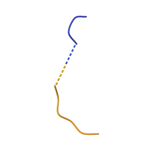
| C-C motif chemokine 3 | 70 | Homo sapiens | Mutation(s): 0 Gene Names: CCL3, G0S19-1, MIP1A, MIP1alpha, SCYA3 | 👁 Image | |
UniProt & NIH Common Fund Data Resources | |||||
Find proteins for P10147 (Homo sapiens) Explore P10147 Go to UniProtKB: P10147 | |||||
PHAROS: P10147 GTEx: ENSG00000277632 | |||||
Entity Groups | |||||
| Sequence Clusters | 30% Identity50% Identity70% Identity90% Identity95% Identity100% Identity | ||||
| UniProt Group | P10147 | ||||
Sequence AnnotationsExpand | |||||
| |||||
| Ligands 2 Unique | |||||
|---|---|---|---|---|---|
| ID | Chains | Name / Formula / InChI Key | 2D Diagram | 3D Interactions | |
| DIO Query on DIO Download Ideal Coordinates CCD File
| E [auth A] F [auth A] G [auth A] I [auth B] J [auth B] E [auth A], F [auth A], G [auth A], I [auth B], J [auth B], K [auth B] | 1,4-DIETHYLENE DIOXIDE C4 H8 O2 RYHBNJHYFVUHQT-UHFFFAOYSA-N | 👁 Image | ||
| ZN Query on ZN Download Ideal Coordinates CCD File | H [auth B], L [auth C] | ZINC ION Zn PTFCDOFLOPIGGS-UHFFFAOYSA-N | 👁 Image | ||
Experimental Data
- Method: X-RAY DIFFRACTION
- Resolution: 3.00 Å
- R-Value Free: 0.237 (Depositor), 0.240 (DCC)
- R-Value Work: 0.183 (Depositor), 0.190 (DCC)
- R-Value Observed: 0.185 (Depositor)
| Length ( Å ) | Angle ( ˚ ) |
|---|---|
| a = 262.737 | α = 90 |
| b = 262.737 | β = 90 |
| c = 90.502 | γ = 120 |
| Software Name | Purpose |
|---|---|
| HKL-2000 | data collection |
| PHASES | phasing |
| REFMAC | refinement |
| HKL-2000 | data reduction |
| SCALEPACK | data scaling |
Deposition Data
- Released Date: 2010-04-14 Deposition Author(s): Ren, M., Guo, Q., Tang, W.J.
Revision History (Full details and data files)
- Version 1.0: 2010-04-14
Type: Initial release - Version 1.1: 2011-07-13
Changes: Version format compliance - Version 1.2: 2021-10-13
Changes: Database references, Derived calculations - Version 1.3: 2023-09-06
Changes: Data collection, Refinement description