
- FASTA Sequence
- PDBx/mmCIF Format
- PDBx/mmCIF Format (gz)
- BinaryCIF Format (gz)
- Legacy PDB Format
- Legacy PDB Format (gz)
- PDBML/XML Format (gz)
- Structure Factors (CIF)
- Structure Factors (CIF - gz)
- Validation Full (PDF - gz)
- Validation (XML - gz)
- Validation (CIF - gz)
- Validation 2fo-fc coefficients (CIF - gz)
- Validation fo-fc coefficients (CIF - gz)
- Biological Assembly 1 (CIF - gz)
- Biological Assembly 1 (PDB - gz)
3IFD | pdb_00003ifd
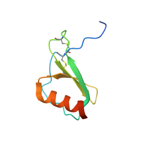
Human synthetic monocyte chemoattractant protein 1 (MCP-1)
- PDB DOI: https://doi.org/10.2210/pdb3IFD/pdb
- Classification: CYTOKINE
- Organism(s): Homo sapiens
- Mutation(s): No
- Deposited: 2009-07-24 Released: 2009-08-04
- Deposition Author(s): Teplyakov, A., Obmolova, G., Gilliland, G.L.
Experimental Data Snapshot
- Method: X-RAY DIFFRACTION
- Resolution: 1.90 Å
- R-Value Free: 0.255 (Depositor), 0.256 (DCC)
- R-Value Work: 0.193 (Depositor), 0.196 (DCC)
- R-Value Observed: 0.195 (Depositor)
Starting Model: experimental
View more details
wwPDB Validation3D Report Full Report
Literature
- 👁 Image
Download Mendeley
Synthesis by native chemical ligation and crystal structure of human CCL2.
Grygiel, T.L., Teplyakov, A., Obmolova, G., Stowell, N., Holland, R., Nemeth, J.F., Pomerantz, S.C., Kruszynski, M., Gilliland, G.L.(2010) Biopolymers 94: 350-359
- PubMed: 20091676 Search on PubMed
- DOI: https://doi.org/10.1002/bip.21390
- Primary Citation Related Structures:
3IFD - PubMed Abstract:
The protein human CC chemokine ligand 2 (CCL2, also known as monocyte chemoattractant protein 1 or MCP-1) has been synthesized using a combination of solid phase peptide synthesis (SPPS) and native chemical ligation (NCL). The thioester-peptide segment was synthesized using the sulfonamide safety-catch linker and 9-fluorenylmethoxycarbonyl (Fmoc) SPPS, and pseudoproline dipeptides were used to facilitate the synthesis of both CCL2 fragments. After assembly of the full-length peptide chain by NCL, a glutathione redox buffer was used to fold and oxidize the CCL2 protein. Synthetic human CCL2 binds to and activates the CCR2 receptor on THP-1 cells, as expected. CCL2 was crystallized and the structure was determined by X-ray diffraction at 1.9-A resolution. The structure of the synthetic protein is very similar to that of a previously reported structure of recombinant human CCL2, although the crystal form is different. The functional CCL2 dimer for the crystal structure reported here is formed around a crystallographic twofold axis. The dimer interface involves residues Val9-Thr10-Cys11, which form an intersubunit antiparallel beta-sheet. Comparison of the CCL2 dimers in different crystal forms indicates a significant flexibility of the quaternary structure. To our knowledge, this is one of the first crystal structures of a protein prepared using the sulfonamide safety-catch linker and NCL.
- Centocor Research and Development, Inc., Radnor, PA 19087, USA. grygieltlr@yahoo.com
Organizational Affiliation:
Explore in 3D: Structure | Sequence Annotations | Electron Density | Validation Report | Ligand Interaction (PO4)
Biological Assembly 1
Explore in 3D: Structure | Sequence Annotations | Electron Density | Validation Report | Ligand Interaction (PO4)
Global Symmetry: Cyclic - C2 (Explore in 3D)
Global Stoichiometry: Homo 2-mer - A2
Find Similar Assemblies
Biological assembly 1 assigned by authors and generated by PISA (software)
Macromolecule Content
- Total Structure Weight: 8.83 kDa
- Atom Count: 554
- Modeled Residue Count: 66
- Deposited Residue Count: 76
- Unique protein chains: 1
Macromolecules
Entity ID: 1 | |||||
|---|---|---|---|---|---|
| Molecule | Chains | Sequence Length | Organism | Details | Image |
| C-C motif chemokine 2 | 76 | Homo sapiens | Mutation(s): 0 | 👁 Image | |
UniProt & NIH Common Fund Data Resources | |||||
PHAROS: P13500 GTEx: ENSG00000108691 | |||||
Entity Groups | |||||
| Sequence Clusters | 30% Identity50% Identity70% Identity90% Identity95% Identity100% Identity | ||||
| UniProt Group | P13500 | ||||
Sequence AnnotationsExpand | |||||
Reference Sequence | |||||
Small Molecules
| Ligands 2 Unique | |||||
|---|---|---|---|---|---|
| ID | Chains | Name / Formula / InChI Key | 2D Diagram | 3D Interactions | |
| PO4 Download:Ideal Coordinates CCD File | C [auth A] | PHOSPHATE ION O4 P NBIIXXVUZAFLBC-UHFFFAOYSA-K | 👁 Image | ||
| K Download:Ideal Coordinates CCD File | B [auth A] | POTASSIUM ION K NPYPAHLBTDXSSS-UHFFFAOYSA-N | 👁 Image | ||
Experimental Data & Validation
Experimental Data
- Method: X-RAY DIFFRACTION
- Resolution: 1.90 Å
- R-Value Free: 0.255 (Depositor), 0.256 (DCC)
- R-Value Work: 0.193 (Depositor), 0.196 (DCC)
- R-Value Observed: 0.195 (Depositor)
| Length ( Å ) | Angle ( ˚ ) |
|---|---|
| a = 60.71 | α = 90 |
| b = 60.71 | β = 90 |
| c = 45.49 | γ = 120 |
| Software Name | Purpose |
|---|---|
| d*TREK | data scaling |
| AMoRE | phasing |
| REFMAC | refinement |
| d*TREK | data reduction |
Entry History
Deposition Data
- Released Date: 2009-08-04 Deposition Author(s): Teplyakov, A., Obmolova, G., Gilliland, G.L.
Revision History (Full details and data files)
- Version 1.0: 2009-08-04
Type: Initial release - Version 1.1: 2011-07-13
Changes: Version format compliance - Version 1.2: 2023-09-06
Changes: Data collection, Database references, Derived calculations, Refinement description - Version 1.3: 2024-11-06
Changes: Structure summary