
- FASTA Sequence
- PDBx/mmCIF Format
- PDBx/mmCIF Format (gz)
- BinaryCIF Format (gz)
- Legacy PDB Format
- Legacy PDB Format (gz)
- PDBML/XML Format (gz)
- Structure Factors (CIF)
- Structure Factors (CIF - gz)
- Validation Full (PDF - gz)
- Validation (XML - gz)
- Validation (CIF - gz)
- Validation 2fo-fc coefficients (CIF - gz)
- Validation fo-fc coefficients (CIF - gz)
- Biological Assembly 1 (CIF - gz)
- Biological Assembly 2 (CIF - gz)
- Biological Assembly 3 (CIF - gz)
- Biological Assembly 1 (PDB - gz)
- Biological Assembly 2 (PDB - gz)
- Biological Assembly 3 (PDB - gz)
3K72 | pdb_00003k72
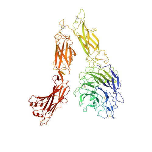
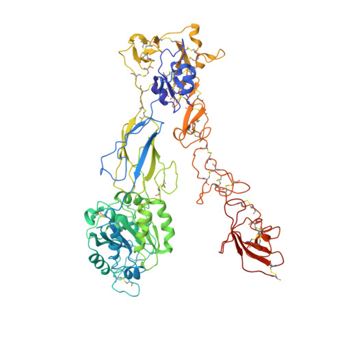
Structure of integrin alphaX beta2
- PDB DOI: https://doi.org/10.2210/pdb3K72/pdb
- Classification: CELL ADHESION
- Organism(s): Homo sapiens
- Expression System: Cricetulus griseus
- Mutation(s): No
- Deposited: 2009-10-11 Released: 2010-01-12
- Deposition Author(s): Xie, C., Zhu, J., Chen, X., Mi, L., Nishida, N., Springer, T.A.
Experimental Data Snapshot
- Method: X-RAY DIFFRACTION
- Resolution: 3.70 Å
- R-Value Free: 0.335 (Depositor), 0.318 (DCC)
- R-Value Work: 0.315 (Depositor), 0.293 (DCC)
- R-Value Observed: 0.315 (Depositor)
Literature
- 👁 Image
Download Mendeley
Structure of an integrin with an alphaI domain, complement receptor type 4.
Xie, C., Zhu, J., Chen, X., Mi, L., Nishida, N., Springer, T.A.(2010) EMBO J 29: 666-679
- PubMed: 20033057 Search on PubMedSearch on PubMed Central
- DOI: https://doi.org/10.1038/emboj.2009.367
- Primary Citation Related Structures:
3K6S, 3K71, 3K72 - PubMed Abstract:
We report the structure of an integrin with an alphaI domain, alpha(X)beta(2), the complement receptor type 4. It was earlier expected that a fixed orientation between the alphaI domain and the beta-propeller domain in which it is inserted would be required for allosteric signal transmission. However, the alphaI domain is highly flexible, enabling two betaI domain conformational states to couple to three alphaI domain states, and greater accessibility for ligand recognition. Although alpha(X)beta(2) is bent similarly to integrins that lack alphaI domains, the terminal domains of the alpha- and beta-legs, calf-2 and beta-tail, are oriented differently than in alphaI-less integrins. Linkers extending to the transmembrane domains are unstructured. Previous mutations in the beta(2)-tail domain support the importance of extension, rather than a deadbolt, in integrin activation. The locations of further activating mutations and antibody epitopes show the critical role of extension, and conversion from the closed to the open headpiece conformation, in integrin activation. Differences among 10 molecules in crystal lattices provide unprecedented information on interdomain flexibility important for modelling integrin extension and activation.
- Department of Pathology, Harvard Medical School, Immune Disease Institute and Children's Hospital, Boston, MA 02115, USA.
Organizational Affiliation:
Explore in 3D: Structure | Sequence Annotations | Electron Density | Validation Report | Ligand Interaction (NAG)
Biological Assembly 1
Explore in 3D: Structure | Sequence Annotations | Electron Density | Validation Report | Ligand Interaction (NAG)
Global Symmetry: Asymmetric - C1
Global Stoichiometry: Hetero 2-mer - A1B1
Find Similar Assemblies
Biological assembly 1 assigned by authors and generated by PISA (software)
Biological Assembly 2
Explore in 3D: Structure | Sequence Annotations | Electron Density | Validation Report | Ligand Interaction (NAG)
Global Symmetry: Asymmetric - C1
Global Stoichiometry: Hetero 2-mer - A1B1
Find Similar Assemblies
Biological assembly 2 assigned by authors and generated by PISA (software)
Biological Assembly 3
Explore in 3D: Structure | Sequence Annotations | Electron Density | Validation Report | Ligand Interaction (NAG)
Global Symmetry: Dihedral - D2 (Explore in 3D)
Global Stoichiometry: Hetero 8-mer - A4B4
Find Similar Assemblies
Biological assembly 3 generated by PISA (software)
Macromolecule Content
- Total Structure Weight: 399.78 kDa
- Atom Count: 24,382
- Modeled Residue Count: 3,112
- Deposited Residue Count: 3,564
- Unique protein chains: 2
Macromolecules
Entity ID: 1 | |||||
|---|---|---|---|---|---|
| Molecule | Chains | Sequence Length | Organism | Details | Image |
| Integrin alpha-X | 1,095 | Homo sapiens | Mutation(s): 0 Gene Names: CD11C, ITGAX | 👁 Image | |
UniProt & NIH Common Fund Data Resources | |||||
PHAROS: P20702 GTEx: ENSG00000140678 | |||||
Entity Groups | |||||
| Sequence Clusters | 30% Identity50% Identity70% Identity90% Identity95% Identity100% Identity | ||||
| UniProt Group | P20702 | ||||
Glycosylation | |||||
| Glycosylation Sites: 5 | Go to GlyGen: P20702-1 | ||||
Sequence AnnotationsExpand | |||||
Reference Sequence | |||||
Entity ID: 2 | |||||
|---|---|---|---|---|---|
| Molecule | Chains | Sequence Length | Organism | Details | Image |
| Integrin beta-2 | 687 | Homo sapiens | Mutation(s): 0 Gene Names: CD18, ITGB2, MFI7 | 👁 Image | |
UniProt & NIH Common Fund Data Resources | |||||
PHAROS: P05107 GTEx: ENSG00000160255 | |||||
Entity Groups | |||||
| Sequence Clusters | 30% Identity50% Identity70% Identity90% Identity95% Identity100% Identity | ||||
| UniProt Group | P05107 | ||||
Glycosylation | |||||
| Glycosylation Sites: 2 | Go to GlyGen: P05107-1 | ||||
Sequence AnnotationsExpand | |||||
Reference Sequence | |||||
Oligosaccharides
HelpEntity ID: 3 | |||||
|---|---|---|---|---|---|
| Molecule | Chains | Length | 2D Diagram | Glycosylation | D Interactions |
| 2-acetamido-2-deoxy-beta-D-glucopyranose-(1-4)-2-acetamido-2-deoxy-beta-D-glucopyranose | E, I | 2 | 👁 Image | N-Glycosylation | |
Glycosylation Resources | |||||
GlyTouCan: G42666HT GlyCosmos: G42666HT GlyGen: G42666HT | |||||
Entity ID: 4 | |||||
|---|---|---|---|---|---|
| Molecule | Chains | Length | 2D Diagram | Glycosylation | D Interactions |
| alpha-D-mannopyranose-(1-3)-[alpha-D-mannopyranose-(1-6)]alpha-D-mannopyranose-(1-4)-2-acetamido-2-deoxy-beta-D-glucopyranose-(1-4)-2-acetamido-2-deoxy-beta-D-glucopyranose | F, J | 5 | 👁 Image | N-Glycosylation | |
Glycosylation Resources | |||||
GlyTouCan: G21381MC GlyCosmos: G21381MC GlyGen: G21381MC | |||||
Small Molecules
Experimental Data & Validation
Experimental Data
- Method: X-RAY DIFFRACTION
- Resolution: 3.70 Å
- R-Value Free: 0.335 (Depositor), 0.318 (DCC)
- R-Value Work: 0.315 (Depositor), 0.293 (DCC)
- R-Value Observed: 0.315 (Depositor)
| Length ( Å ) | Angle ( ˚ ) |
|---|---|
| a = 160.962 | α = 90 |
| b = 165.549 | β = 90 |
| c = 536.178 | γ = 90 |
| Software Name | Purpose |
|---|---|
| MAR345dtb | data collection |
| SOLVE | phasing |
| PHENIX | refinement |
| XDS | data reduction |
| XDS | data scaling |
Entry History
Deposition Data
- Released Date: 2010-01-12 Deposition Author(s): Xie, C., Zhu, J., Chen, X., Mi, L., Nishida, N., Springer, T.A.
Revision History (Full details and data files)
- Version 1.0: 2010-01-12
Type: Initial release - Version 1.1: 2011-07-13
Changes: Version format compliance - Version 2.0: 2020-07-29
Type: Remediation
Reason: Carbohydrate remediation
Changes: Advisory, Atomic model, Data collection, Database references, Derived calculations, Structure summary - Version 2.1: 2024-11-27
Changes: Data collection, Database references, Structure summary