
- FASTA Sequence
- PDBx/mmCIF Format
- PDBx/mmCIF Format (gz)
- BinaryCIF Format (gz)
- Legacy PDB Format
- Legacy PDB Format (gz)
- PDBML/XML Format (gz)
- Structure Factors (CIF)
- Structure Factors (CIF - gz)
- Validation Full (PDF - gz)
- Validation (XML - gz)
- Validation (CIF - gz)
- Validation 2fo-fc coefficients (CIF - gz)
- Validation fo-fc coefficients (CIF - gz)
- Biological Assembly 1 (CIF - gz)
- Biological Assembly 2 (CIF - gz)
- Biological Assembly 3 (CIF - gz)
- Biological Assembly 1 (PDB - gz)
- Biological Assembly 2 (PDB - gz)
- Biological Assembly 3 (PDB - gz)
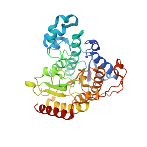
3MAX | pdb_00003max
Crystal Structure of Human HDAC2 complexed with an N-(2-aminophenyl)benzamide
- PDB DOI: https://doi.org/10.2210/pdb3MAX/pdb
- Classification: HYDROLASE
- Organism(s): Homo sapiens
- Expression System: unidentified baculovirus
- Mutation(s): No
- Deposited: 2010-03-24 Released: 2010-04-28
- Deposition Author(s): Skene, R.J., Jennings, A.J.
Experimental Data Snapshot
- Method: X-RAY DIFFRACTION
- Resolution: 2.05 Å
- R-Value Free: 0.204 (Depositor), 0.220 (DCC)
- R-Value Work: 0.164 (Depositor), 0.180 (DCC)
- R-Value Observed: 0.166 (Depositor)
- 👁 Image
Download Mendeley
Exploration of the HDAC2 foot pocket: Synthesis and SAR of substituted N-(2-aminophenyl)benzamides.
Bressi, J.C., Jennings, A.J., Skene, R., Wu, Y., Melkus, R., De Jong, R., O'Connell, S., Grimshaw, C.E., Navre, M., Gangloff, A.R.(2010) Bioorg Med Chem Lett 20: 3142-3145
- PubMed: 20392638 Search on PubMed
- DOI: https://doi.org/10.1016/j.bmcl.2010.03.091
- Primary Citation Related Structures:
3MAX - PubMed Abstract:
A series of N-(2-amino-5-substituted phenyl)benzamides (3-21) were designed, synthesized and evaluated for their inhibition of HDAC2 and their cytotoxicity in HCT116 cancer cells. Multiple compounds from this series demonstrated time-dependent binding kinetics that is rationalized using a co-complex crystal structure of HDAC2 and N-(4-aminobiphenyl-3-yl)benzamide (6).
- Takeda San Diego, San Diego, CA 92121, USA.
Organizational Affiliation:
Explore in 3D: Structure | Sequence Annotations | Electron Density | Validation Report | Ligand Interaction (LLX)
Explore in 3D: Structure | Sequence Annotations | Electron Density | Validation Report | Ligand Interaction (LLX)
Global Symmetry: Asymmetric - C1
Global Stoichiometry: Monomer - A1
Find Similar Assemblies
Biological assembly 1 assigned by authors and generated by PISA (software)
Explore in 3D: Structure | Sequence Annotations | Electron Density | Validation Report | Ligand Interaction (LLX)
Global Symmetry: Asymmetric - C1
Global Stoichiometry: Monomer - A1
Find Similar Assemblies
Biological assembly 2 assigned by authors and generated by PISA (software)
Explore in 3D: Structure | Sequence Annotations | Electron Density | Validation Report | Ligand Interaction (LLX)
Global Symmetry: Asymmetric - C1
Global Stoichiometry: Monomer - A1
Find Similar Assemblies
Biological assembly 3 assigned by authors and generated by PISA (software)
Macromolecule Content
- Total Structure Weight: 127.9 kDa
- Atom Count: 9,584
- Modeled Residue Count: 1,097
- Deposited Residue Count: 1,101
- Unique protein chains: 1
Entity ID: 1 | |||||
|---|---|---|---|---|---|
| Molecule | Chains | Sequence Length | Organism | Details | Image |
| Histone deacetylase 2 | 367 | Homo sapiens | Mutation(s): 0 Gene Names: HDAC2 EC: 3.5.1.98 (PDB Primary Data), 3.5.1 (UniProt) | 👁 Image | |
UniProt & NIH Common Fund Data Resources | |||||
Find proteins for Q92769 (Homo sapiens) Explore Q92769 Go to UniProtKB: Q92769 | |||||
PHAROS: Q92769 GTEx: ENSG00000196591 | |||||
Entity Groups | |||||
| Sequence Clusters | 30% Identity50% Identity70% Identity90% Identity95% Identity100% Identity | ||||
| UniProt Group | Q92769 | ||||
Sequence AnnotationsExpand | |||||
| |||||
| Ligands 5 Unique | |||||
|---|---|---|---|---|---|
| ID | Chains | Name / Formula / InChI Key | 2D Diagram | 3D Interactions | |
| LLX Query on LLX Download Ideal Coordinates CCD File | G [auth A], L [auth B], P [auth C] | N-(4-aminobiphenyl-3-yl)benzamide C19 H16 N2 O ZWLFHHHQRUYIBT-UHFFFAOYSA-N | 👁 Image | ||
| NHE Query on NHE Download Ideal Coordinates CCD File | H [auth A] | 2-[N-CYCLOHEXYLAMINO]ETHANE SULFONIC ACID C8 H17 N O3 S MKWKNSIESPFAQN-UHFFFAOYSA-N | 👁 Image | ||
| ZN Query on ZN Download Ideal Coordinates CCD File | D [auth A], I [auth B], M [auth C] | ZINC ION Zn PTFCDOFLOPIGGS-UHFFFAOYSA-N | 👁 Image | ||
| CA Query on CA Download Ideal Coordinates CCD File | E [auth A], J [auth B], N [auth C] | CALCIUM ION Ca BHPQYMZQTOCNFJ-UHFFFAOYSA-N | 👁 Image | ||
| NA Query on NA Download Ideal Coordinates CCD File | F [auth A], K [auth B], O [auth C] | SODIUM ION Na FKNQFGJONOIPTF-UHFFFAOYSA-N | 👁 Image | ||
Experimental Data
- Method: X-RAY DIFFRACTION
- Resolution: 2.05 Å
- R-Value Free: 0.204 (Depositor), 0.220 (DCC)
- R-Value Work: 0.164 (Depositor), 0.180 (DCC)
- R-Value Observed: 0.166 (Depositor)
| Length ( Å ) | Angle ( ˚ ) |
|---|---|
| a = 92.311 | α = 90 |
| b = 97.227 | β = 90 |
| c = 138.849 | γ = 90 |
| Software Name | Purpose |
|---|---|
| HKL-2000 | data collection |
| MOLREP | phasing |
| REFMAC | refinement |
| HKL-2000 | data reduction |
| HKL-2000 | data scaling |
Deposition Data
- Released Date: 2010-04-28 Deposition Author(s): Skene, R.J., Jennings, A.J.
Revision History (Full details and data files)
- Version 1.0: 2010-04-28
Type: Initial release - Version 1.1: 2011-07-13
Changes: Version format compliance - Version 1.2: 2024-02-21
Changes: Data collection, Database references, Derived calculations