
- FASTA Sequence
- PDBx/mmCIF Format
- PDBx/mmCIF Format (gz)
- BinaryCIF Format (gz)
- Legacy PDB Format
- Legacy PDB Format (gz)
- PDBML/XML Format (gz)
- Structure Factors (CIF)
- Structure Factors (CIF - gz)
- Validation Full (PDF - gz)
- Validation (XML - gz)
- Validation (CIF - gz)
- Validation 2fo-fc coefficients (CIF - gz)
- Validation fo-fc coefficients (CIF - gz)
- Biological Assembly 1 (CIF - gz)
- Biological Assembly 2 (CIF - gz)
- Biological Assembly 1 (PDB - gz)
- Biological Assembly 2 (PDB - gz)
3NID | pdb_00003nid
The Closed Headpiece of Integrin alphaIIB beta3 and its Complex with an alpahIIB beta3 -Specific Antagonist That Does Not Induce Opening
- PDB DOI: https://doi.org/10.2210/pdb3NID/pdb
- Classification: CELL ADHESION/BLOOD CLOTTING
- Organism(s): Homo sapiens, Mus musculus
- Expression System: Cricetulus griseus
- Mutation(s): No
- Deposited: 2010-06-15 Released: 2010-12-22
- Deposition Author(s): Zhu, J.H., Zhu, J.Q., Springer, T.A.
Experimental Data Snapshot
- Method: X-RAY DIFFRACTION
- Resolution: 2.30 Å
- R-Value Free: 0.215 (Depositor), 0.231 (DCC)
- R-Value Work: 0.179 (Depositor), 0.192 (DCC)
- R-Value Observed: 0.179 (Depositor)
Starting Model: experimental
View more details
Literature
- 👁 Image
Download Mendeley
Closed headpiece of integrin {alpha}IIb{beta}3 and its complex with an {alpha}IIb{beta}3-specific antagonist that does not induce opening.
Zhu, J.H., Zhu, J.Q., Negri, A., Provasi, D., Filizola, M., Coller, B.S., Springer, T.A.(2010) Blood 116: 5050-5059
- PubMed: 20679525 Search on PubMedSearch on PubMed Central
- DOI: https://doi.org/10.1182/blood-2010-04-281154
- Primary Citation Related Structures:
3NID, 3NIF, 3NIG - PubMed Abstract:
The platelet integrin α(IIb)β(3) is essential for hemostasis and thrombosis through its binding of adhesive plasma proteins. We have determined crystal structures of the α(IIb)β(3) headpiece in the absence of ligand and after soaking in RUC-1, a novel small molecule antagonist. In the absence of ligand, the α(IIb)β(3) headpiece is in a closed conformation, distinct from the open conformation visualized in presence of Arg-Gly-Asp (RGD) antagonists. In contrast to RGD antagonists, RUC-1 binds only to the α(IIb) subunit. Molecular dynamics revealed nearly identical binding. Two species-specific residues, α(IIb) Y190 and α(IIb) D232, in the RUC-1 binding site were confirmed as important by mutagenesis. In sharp contrast to RGD-based antagonists, RUC-1 did not induce α(IIb)β(3) to adopt an open conformation, as determined by gel filtration and dynamic light scattering. These studies provide insights into the factors that regulate integrin headpiece opening, and demonstrate the molecular basis for a novel mechanism of integrin antagonism.
- Immune Disease Institute, Children's Hospital Boston, and Department of Pathology, Harvard Medical School, Boston, MA 02115, USA.
Organizational Affiliation:
Explore in 3D: Structure | Sequence Annotations | Electron Density | Validation Report | Ligand Interaction (NAG)
Biological Assembly 1
Explore in 3D: Structure | Sequence Annotations | Electron Density | Validation Report | Ligand Interaction (NAG)
Global Symmetry: Asymmetric - C1
Global Stoichiometry: Hetero 4-mer - A1B1C1D1
Pseudo Symmetry: Asymmetric - C1
Pseudo Stoichiometry: Hetero 4-mer - A2B1C1
Find Similar Assemblies
Biological assembly 1 assigned by authors.
Biological Assembly 2
Explore in 3D: Structure | Sequence Annotations | Electron Density | Validation Report | Ligand Interaction (NAG)
Global Symmetry: Asymmetric - C1
Global Stoichiometry: Hetero 4-mer - A1B1C1D1
Pseudo Symmetry: Asymmetric - C1
Pseudo Stoichiometry: Hetero 4-mer - A2B1C1
Find Similar Assemblies
Biological assembly 2 assigned by authors.
Macromolecule Content
- Total Structure Weight: 302.26 kDa
- Atom Count: 22,131
- Modeled Residue Count: 2,702
- Deposited Residue Count: 2,726
- Unique protein chains: 4
Macromolecules
Entity ID: 1 | |||||
|---|---|---|---|---|---|
| Molecule | Chains | Sequence Length | Organism | Details | Image |
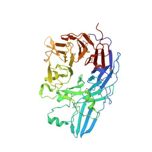
| Integrin alpha-IIb | 457 | Homo sapiens | Mutation(s): 0 Gene Names: ITGA2B, GP2B, ITGAB | 👁 Image | |
UniProt & NIH Common Fund Data Resources | |||||
PHAROS: P08514 GTEx: ENSG00000005961 | |||||
Entity Groups | |||||
| Sequence Clusters | 30% Identity50% Identity70% Identity90% Identity95% Identity100% Identity | ||||
| UniProt Group | P08514 | ||||
Sequence AnnotationsExpand | |||||
Reference Sequence | |||||
Entity ID: 2 | |||||
|---|---|---|---|---|---|
| Molecule | Chains | Sequence Length | Organism | Details | Image |
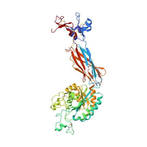
| Integrin beta-3 | 471 | Homo sapiens | Mutation(s): 0 Gene Names: ITGB3, GP3A | 👁 Image | |
UniProt & NIH Common Fund Data Resources | |||||
PHAROS: P05106 GTEx: ENSG00000259207 | |||||
Entity Groups | |||||
| Sequence Clusters | 30% Identity50% Identity70% Identity90% Identity95% Identity100% Identity | ||||
| UniProt Group | P05106 | ||||
Glycosylation | |||||
| Glycosylation Sites: 3 | Go to GlyGen: P05106-1 | ||||
Sequence AnnotationsExpand | |||||
Reference Sequence | |||||
Entity ID: 3 | |||||
|---|---|---|---|---|---|
| Molecule | Chains | Sequence Length | Organism | Details | Image |
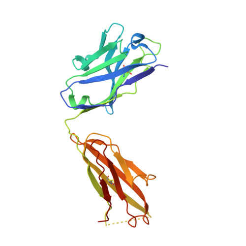
| Monoclonal antibody 10E5 heavy chain | E, G [auth H] | 221 | Mus musculus | Mutation(s): 0 | 👁 Image |
Entity ID: 4 | |||||
|---|---|---|---|---|---|
| Molecule | Chains | Sequence Length | Organism | Details | Image |
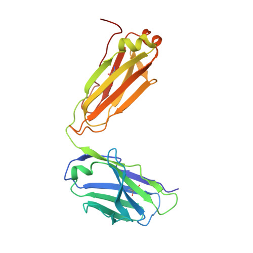
| Monoclonal antibody 10E5 light chain | F, H [auth L] | 214 | Mus musculus | Mutation(s): 0 | 👁 Image |
Oligosaccharides
HelpEntity ID: 5 | |||||
|---|---|---|---|---|---|
| Molecule | Chains | Length | 2D Diagram | Glycosylation | D Interactions |
| alpha-D-mannopyranose-(1-3)-[alpha-D-mannopyranose-(1-6)]alpha-D-mannopyranose-(1-4)-2-acetamido-2-deoxy-beta-D-glucopyranose-(1-4)-2-acetamido-2-deoxy-beta-D-glucopyranose | I [auth G] | 5 | 👁 Image | N-Glycosylation | |
Glycosylation Resources | |||||
GlyTouCan: G21381MC GlyCosmos: G21381MC GlyGen: G21381MC | |||||
Entity ID: 6 | |||||
|---|---|---|---|---|---|
| Molecule | Chains | Length | 2D Diagram | Glycosylation | D Interactions |
| 2-acetamido-2-deoxy-beta-D-glucopyranose-(1-4)-2-acetamido-2-deoxy-beta-D-glucopyranose | J [auth I], L [auth K] | 2 | 👁 Image | N-Glycosylation | |
Glycosylation Resources | |||||
GlyTouCan: G42666HT GlyCosmos: G42666HT GlyGen: G42666HT | |||||
Small Molecules
Experimental Data & Validation
Experimental Data
- Method: X-RAY DIFFRACTION
- Resolution: 2.30 Å
- R-Value Free: 0.215 (Depositor), 0.231 (DCC)
- R-Value Work: 0.179 (Depositor), 0.192 (DCC)
- R-Value Observed: 0.179 (Depositor)
| Length ( Å ) | Angle ( ˚ ) |
|---|---|
| a = 258.97 | α = 90 |
| b = 144.49 | β = 90 |
| c = 104.2 | γ = 90 |
| Software Name | Purpose |
|---|---|
| Blu-Ice | data collection |
| PHASER | phasing |
| REFMAC | refinement |
| XDS | data reduction |
| XDS | data scaling |
Entry History
Deposition Data
- Released Date: 2010-12-22 Deposition Author(s): Zhu, J.H., Zhu, J.Q., Springer, T.A.
Revision History (Full details and data files)
- Version 1.0: 2010-12-22
Type: Initial release - Version 1.1: 2011-07-13
Changes: Version format compliance - Version 2.0: 2020-07-29
Type: Remediation
Reason: Carbohydrate remediation
Changes: Advisory, Atomic model, Data collection, Derived calculations, Structure summary - Version 2.1: 2023-09-06
Changes: Advisory, Data collection, Database references, Refinement description, Structure summary - Version 2.2: 2024-11-06
Changes: Structure summary