
- FASTA Sequence
- PDBx/mmCIF Format
- PDBx/mmCIF Format (gz)
- BinaryCIF Format (gz)
- Legacy PDB Format
- Legacy PDB Format (gz)
- PDBML/XML Format (gz)
- Structure Factors (CIF)
- Structure Factors (CIF - gz)
- Validation Full (PDF - gz)
- Validation (XML - gz)
- Validation (CIF - gz)
- Validation 2fo-fc coefficients (CIF - gz)
- Validation fo-fc coefficients (CIF - gz)
- Biological Assembly 1 (CIF - gz)
- Biological Assembly 1 (PDB - gz)
3REQ | pdb_00003req
METHYLMALONYL-COA MUTASE, SUBSTRATE-FREE STATE (POOR QUALITY STRUCTURE)
- PDB DOI: https://doi.org/10.2210/pdb3REQ/pdb
- Classification: COMPLEX (ISOMERASE/DEOXYADENOSINE)
- Organism(s): Propionibacterium freudenreichii subsp. shermanii
- Expression System: Escherichia coli
- Mutation(s): No
- Deposited: 1997-12-04 Released: 1998-03-18
- Deposition Author(s): Evans, P.R., Mancia, F.
Experimental Data Snapshot
- Method: X-RAY DIFFRACTION
- Resolution: 2.70 Å
- R-Value Free: 0.393 (Depositor), 0.350 (DCC)
- R-Value Work: 0.313 (Depositor), 0.280 (DCC)
Starting Model: experimental
View more details
- 👁 Image
Download Mendeley
Conformational changes on substrate binding to methylmalonyl CoA mutase and new insights into the free radical mechanism.
Mancia, F., Evans, P.R.(1998) Structure 6: 711-720
- PubMed: 9655823 Search on PubMed
- DOI: https://doi.org/10.1016/s0969-2126(98)00073-2
- Primary Citation Related Structures:
2REQ, 3REQ, 4REQ - PubMed Abstract:
Methylmalonyl CoA mutase catalyses the interconversion of succinyl CoA and methylmalonyl CoA via a free radical mechanism. The enzyme belongs to a family of enzymes that catalyse intramolecular rearrangement reactions in which a group and a hydrogen atom on adjacent carbons are exchanged. These enzymes use the cofactor adenosylcobalamin (coenzyme B12) which breaks to form an adenosyl radical, thus initiating the reaction. Determination of the structure of substrate-free methylmalonyl CoA mutase was initiated to provide further insight into the mechanism of radical formation. We report here two structures of methylmalonyl CoA mutase from Propionibacterium shermanii. The first structure is of the enzyme in a nonproductive complex with CoA at 2.5 A resolution. This structure serves as a model for the substrate-free conformation of the enzyme, as it is very similar to the second much poorer 2.7 A resolution structure derived from a truly substrate-free crystal. The true substrate-free structure also shows the adenosyl group bound to the cobalt atom. Comparison of this structure with that of the previously reported complex of the enzyme with a substrate analogue shows that major conformational changes occur upon substrate binding. The substrate-binding site of the enzyme is located within a (beta alpha)8 TIM-barrel domain. In the absence of substrate, this TIM-barrel domain is split apart and the active site is accessible to solvent. When substrate binds, the barrel closes up with the substrate along its axis and the active site becomes completely buried. The closure of the active-site cavity upon substrate binding displaces the adenosyl group of the cofactor from the central cobalt atom into the active-site cavity. This triggers the formation of the free radical that initiates the rearrangement reaction. The TIM-barrel domain is substantially different from all others yet reported: in its unliganded form it is broken open, exposing the small hydrophilic sidechains which fill the centre. The typical barrel structure is only formed when substrate is bound.
- MRC Laboratory of Molecular Biology Hills Road, Cambridge, CB2 2QH, UK.
Organizational Affiliation:
Explore in 3D: Structure | Sequence Annotations | Electron Density | Validation Report | Ligand Interaction (B12)
Explore in 3D: Structure | Sequence Annotations | Electron Density | Validation Report | Ligand Interaction (B12)
Global Symmetry: Asymmetric - C1
Global Stoichiometry: Hetero 2-mer - A1B1
Find Similar Assemblies
Biological assembly 1 assigned by authors and generated by PISA (software)
Macromolecule Content
- Total Structure Weight: 151.17 kDa
- Atom Count: 10,285
- Modeled Residue Count: 1,345
- Deposited Residue Count: 1,364
- Unique protein chains: 2
Entity ID: 1 | |||||
|---|---|---|---|---|---|
| Molecule | Chains | Sequence Length | Organism | Details | Image |
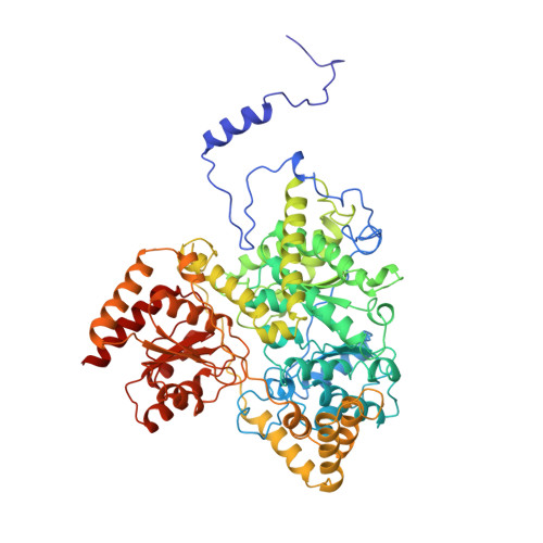
| METHYLMALONYL-COA MUTASE | 727 | Propionibacterium freudenreichii subsp. shermanii | Mutation(s): 0 Gene Names: MUTA, MUTB EC: 5.4.99.2 | 👁 Image | |
UniProt | |||||
Find proteins for P11653 (Propionibacterium freudenreichii subsp. shermanii) Explore P11653 Go to UniProtKB: P11653 | |||||
Entity Groups | |||||
| Sequence Clusters | 30% Identity50% Identity70% Identity90% Identity95% Identity100% Identity | ||||
| UniProt Group | P11653 | ||||
Sequence AnnotationsExpand | |||||
| |||||
Entity ID: 2 | |||||
|---|---|---|---|---|---|
| Molecule | Chains | Sequence Length | Organism | Details | Image |
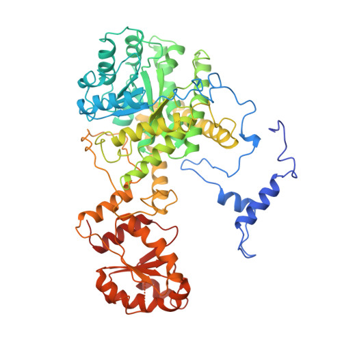
| METHYLMALONYL-COA MUTASE | 637 | Propionibacterium freudenreichii subsp. shermanii | Mutation(s): 0 Gene Names: MUTA, MUTB EC: 5.4.99.2 | 👁 Image | |
UniProt | |||||
Find proteins for P11652 (Propionibacterium freudenreichii subsp. shermanii) Explore P11652 Go to UniProtKB: P11652 | |||||
Entity Groups | |||||
| Sequence Clusters | 30% Identity50% Identity70% Identity90% Identity95% Identity100% Identity | ||||
| UniProt Group | P11652 | ||||
Sequence AnnotationsExpand | |||||
| |||||
| Ligands 2 Unique | |||||
|---|---|---|---|---|---|
| ID | Chains | Name / Formula / InChI Key | 2D Diagram | 3D Interactions | |
| B12 Query on B12 Download Ideal Coordinates CCD File | C [auth A] | COBALAMIN C62 H89 Co N13 O14 P LKVIQTCSMMVGFU-DWSMJLPVSA-N | 👁 Image | ||
| ADN Query on ADN Download Ideal Coordinates CCD File | D [auth A] | ADENOSINE C10 H13 N5 O4 OIRDTQYFTABQOQ-KQYNXXCUSA-N | 👁 Image | ||
Experimental Data
- Method: X-RAY DIFFRACTION
- Resolution: 2.70 Å
- R-Value Free: 0.393 (Depositor), 0.350 (DCC)
- R-Value Work: 0.313 (Depositor), 0.280 (DCC)
| Length ( Å ) | Angle ( ˚ ) |
|---|---|
| a = 110.91 | α = 90 |
| b = 110.91 | β = 90 |
| c = 257.74 | γ = 90 |
| Software Name | Purpose |
|---|---|
| MOSFLM | data reduction |
| SCALA | data scaling |
| AMoRE | phasing |
| REFMAC | refinement |
| CCP4 | data scaling |
Deposition Data
- Released Date: 1998-03-18 Deposition Author(s): Evans, P.R., Mancia, F.
Revision History (Full details and data files)
- Version 1.0: 1998-03-18
Type: Initial release - Version 1.1: 2008-03-25
Changes: Version format compliance - Version 1.2: 2011-07-13
Changes: Version format compliance - Version 1.3: 2012-10-24
Changes: Non-polymer description - Version 1.4: 2023-08-09
Changes: Database references, Derived calculations, Refinement description - Version 1.5: 2024-05-22
Changes: Data collection