
- FASTA Sequence
- PDBx/mmCIF Format
- PDBx/mmCIF Format (gz)
- BinaryCIF Format (gz)
- Legacy PDB Format
- Legacy PDB Format (gz)
- PDBML/XML Format (gz)
- Structure Factors (CIF)
- Structure Factors (CIF - gz)
- Validation Full (PDF - gz)
- Validation (XML - gz)
- Validation (CIF - gz)
- Validation 2fo-fc coefficients (CIF - gz)
- Validation fo-fc coefficients (CIF - gz)
- Biological Assembly 1 (CIF - gz)
- Biological Assembly 2 (CIF - gz)
- Biological Assembly 3 (CIF - gz)
- Biological Assembly 1 (PDB - gz)
- Biological Assembly 2 (PDB - gz)
- Biological Assembly 3 (PDB - gz)
3RPG | pdb_00003rpg
Bmi1/Ring1b-UbcH5c complex structure
- PDB DOI: https://doi.org/10.2210/pdb3RPG/pdb
- Classification: LIGASE
- Organism(s): Homo sapiens
- Expression System: Escherichia coli
- Mutation(s): No
- Deposited: 2011-04-26 Released: 2011-08-17
- Deposition Author(s): Bentley, M.L., Dong, K.C., Cochran, A.G.
Experimental Data Snapshot
- Method: X-RAY DIFFRACTION
- Resolution: 2.65 Å
- R-Value Free: 0.243 (Depositor), 0.240 (DCC)
- R-Value Work: 0.217 (Depositor), 0.210 (DCC)
- R-Value Observed: 0.219 (Depositor)
Starting Models: experimental
View more details
wwPDB Validation 3D Report Full Report
- 👁 Image
Download Mendeley
Recognition of UbcH5c and the nucleosome by the Bmi1/Ring1b ubiquitin ligase complex.
Bentley, M.L., Corn, J.E., Dong, K.C., Phung, Q., Cheung, T.K., Cochran, A.G.(2011) EMBO J 30: 3285-3297
- PubMed: 21772249 Search on PubMedSearch on PubMed Central
- DOI: https://doi.org/10.1038/emboj.2011.243
- Primary Citation Related Structures:
3RPG - PubMed Abstract:
The Polycomb repressive complex 1 (PRC1) mediates gene silencing, in part by monoubiquitination of histone H2A on lysine 119 (uH2A). Bmi1 and Ring1b are critical components of PRC1 that heterodimerize via their N-terminal RING domains to form an active E3 ubiquitin ligase. We have determined the crystal structure of a complex between the Bmi1/Ring1b RING-RING heterodimer and the E2 enzyme UbcH5c and find that UbcH5c interacts with Ring1b only, in a manner fairly typical of E2-E3 interactions. However, we further show that the Bmi1/Ring1b RING domains bind directly to duplex DNA through a basic surface patch unique to the Bmi1/Ring1b RING-RING dimer. Mutation of residues on this interaction surface leads to a loss of H2A ubiquitination activity. Computational modelling of the interface between Bmi1/Ring1b-UbcH5c and the nucleosome suggests that Bmi1/Ring1b interacts with both nucleosomal DNA and an acidic patch on histone H4 to achieve specific monoubiquitination of H2A. Our results point to a novel mechanism of substrate recognition, and control of product formation, by Bmi1/Ring1b.
- Department of Early Discovery Biochemistry, Genentech Research and Early Development, South San Francisco, CA, USA.
Organizational Affiliation:
Explore in 3D: Structure | Sequence Annotations | Electron Density | Validation Report | Ligand Interaction (ZN)
Explore in 3D: Structure | Sequence Annotations | Electron Density | Validation Report | Ligand Interaction (ZN)
Global Symmetry: Asymmetric - C1
Global Stoichiometry: Hetero 3-mer - A1B1C1
Pseudo Symmetry: Asymmetric - C1
Pseudo Stoichiometry: Hetero 3-mer - A2B1
Find Similar Assemblies
Biological assembly 1 assigned by authors.
Explore in 3D: Structure | Sequence Annotations | Electron Density | Validation Report | Ligand Interaction (ZN)
Global Symmetry: Asymmetric - C1
Global Stoichiometry: Monomer - A1
Find Similar Assemblies
Biological assembly 2 generated by PISA (software)
Explore in 3D: Structure | Sequence Annotations | Electron Density | Validation Report | Ligand Interaction (ZN)
Global Symmetry: Asymmetric - C1
Global Stoichiometry: Hetero 2-mer - A1B1
Pseudo Symmetry: Cyclic - C2 (Explore in 3D)
Pseudo Stoichiometry: Homo 2-mer - A2
Find Similar Assemblies
Biological assembly 3 generated by PISA (software)
Macromolecule Content
- Total Structure Weight: 44.26 kDa
- Atom Count: 2,912
- Modeled Residue Count: 349
- Deposited Residue Count: 387
- Unique protein chains: 3
Entity ID: 1 | |||||
|---|---|---|---|---|---|
| Molecule | Chains | Sequence Length | Organism | Details | Image |
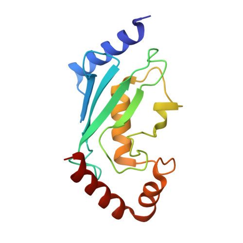
| Ubiquitin-conjugating enzyme E2 D3 | 149 | Homo sapiens | Mutation(s): 0 Gene Names: UBE2D3, UBC5C, UBCH5C EC: 6.3.2.19 (PDB Primary Data), 2.3.2.24 (UniProt), 2.3.2.23 (UniProt) | 👁 Image | |
UniProt & NIH Common Fund Data Resources | |||||
Find proteins for P61077 (Homo sapiens) Explore P61077 Go to UniProtKB: P61077 | |||||
PHAROS: P61077 GTEx: ENSG00000109332 | |||||
Entity Groups | |||||
| Sequence Clusters | 30% Identity50% Identity70% Identity90% Identity95% Identity100% Identity | ||||
| UniProt Group | P61077 | ||||
Sequence AnnotationsExpand | |||||
| |||||
Entity ID: 2 | |||||
|---|---|---|---|---|---|
| Molecule | Chains | Sequence Length | Organism | Details | Image |
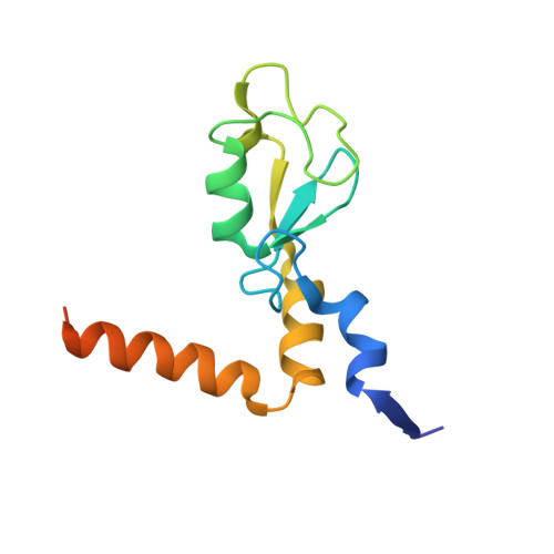
| Polycomb complex protein BMI-1 | 117 | Homo sapiens | Mutation(s): 0 Gene Names: BMI1, PCGF4, RNF51 | 👁 Image | |
UniProt & NIH Common Fund Data Resources | |||||
Find proteins for P35226 (Homo sapiens) Explore P35226 Go to UniProtKB: P35226 | |||||
PHAROS: P35226 GTEx: ENSG00000168283 | |||||
Entity Groups | |||||
| Sequence Clusters | 30% Identity50% Identity70% Identity90% Identity95% Identity100% Identity | ||||
| UniProt Group | P35226 | ||||
Sequence AnnotationsExpand | |||||
| |||||
Entity ID: 3 | |||||
|---|---|---|---|---|---|
| Molecule | Chains | Sequence Length | Organism | Details | Image |
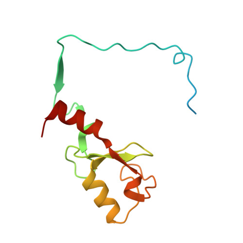
| E3 ubiquitin-protein ligase RING2 | 121 | Homo sapiens | Mutation(s): 0 Gene Names: RNF2, BAP1, DING, HIPI3, RING1B EC: 6.3.2 (PDB Primary Data), 2.3.2.27 (UniProt) | 👁 Image | |
UniProt & NIH Common Fund Data Resources | |||||
Find proteins for Q99496 (Homo sapiens) Explore Q99496 Go to UniProtKB: Q99496 | |||||
PHAROS: Q99496 GTEx: ENSG00000121481 | |||||
Entity Groups | |||||
| Sequence Clusters | 30% Identity50% Identity70% Identity90% Identity95% Identity100% Identity | ||||
| UniProt Group | Q99496 | ||||
Sequence AnnotationsExpand | |||||
| |||||
| Ligands 1 Unique | |||||
|---|---|---|---|---|---|
| ID | Chains | Name / Formula / InChI Key | 2D Diagram | 3D Interactions | |
| ZN Query on ZN Download Ideal Coordinates CCD File
| D [auth B], E [auth B], F [auth C], G [auth C] | ZINC ION Zn PTFCDOFLOPIGGS-UHFFFAOYSA-N | 👁 Image | ||
Experimental Data
- Method: X-RAY DIFFRACTION
- Resolution: 2.65 Å
- R-Value Free: 0.243 (Depositor), 0.240 (DCC)
- R-Value Work: 0.217 (Depositor), 0.210 (DCC)
- R-Value Observed: 0.219 (Depositor)
| Length ( Å ) | Angle ( ˚ ) |
|---|---|
| a = 107.89 | α = 90 |
| b = 107.89 | β = 90 |
| c = 77.572 | γ = 120 |
| Software Name | Purpose |
|---|---|
| StructureStudio | data collection |
| PHASER | phasing |
| PHENIX | refinement |
| HKL-2000 | data reduction |
| HKL-2000 | data scaling |
Deposition Data
- Released Date: 2011-08-17 Deposition Author(s): Bentley, M.L., Dong, K.C., Cochran, A.G.
Revision History (Full details and data files)
- Version 1.0: 2011-08-17
Type: Initial release - Version 1.1: 2011-08-31
Changes: Database references - Version 1.2: 2023-09-13
Changes: Data collection, Database references, Derived calculations, Refinement description