
- FASTA Sequence
- PDBx/mmCIF Format
- PDBx/mmCIF Format (gz)
- BinaryCIF Format (gz)
- Legacy PDB Format
- Legacy PDB Format (gz)
- PDBML/XML Format (gz)
- Structure Factors (CIF)
- Structure Factors (CIF - gz)
- Validation Full (PDF - gz)
- Validation (XML - gz)
- Validation (CIF - gz)
- Validation 2fo-fc coefficients (CIF - gz)
- Validation fo-fc coefficients (CIF - gz)
- Biological Assembly 1 (CIF - gz)
- Biological Assembly 1 (PDB - gz)
3UJE | pdb_00003uje
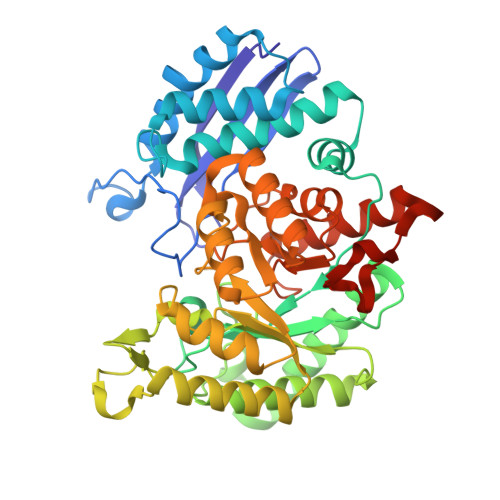
Asymmetric complex of human neuron specific enolase-3-PGA/PEP
- PDB DOI: https://doi.org/10.2210/pdb3UJE/pdb
- Classification: LYASE
- Organism(s): Homo sapiens
- Expression System: Escherichia coli
- Mutation(s): No
- Deposited: 2011-11-07 Released: 2012-08-22
- Deposition Author(s): Qin, J., Chai, G., Brewer, J., Lovelace, L., Lebioda, L.
Experimental Data Snapshot
- Method: X-RAY DIFFRACTION
- Resolution: 1.55 Å
- R-Value Free: 0.191 (Depositor), 0.190 (DCC)
- R-Value Work: 0.146 (Depositor), 0.140 (DCC)
- R-Value Observed: 0.148 (Depositor)
Starting Model: experimental
View more details
- 👁 Image
Download Mendeley
Structures of asymmetric complexes of human neuron specific enolase with resolved substrate and product and an analogous complex with two inhibitors indicate subunit interaction and inhibitor cooperativity.
Qin, J., Chai, G., Brewer, J.M., Lovelace, L.L., Lebioda, L.(2012) J Inorg Biochem 111: 187-194
- PubMed: 22437160 Search on PubMedSearch on PubMed Central
- DOI: https://doi.org/10.1016/j.jinorgbio.2012.02.011
- Primary Citation Related Structures:
3UCC, 3UCD, 3UJE, 3UJF, 3UJR, 3UJS - PubMed Abstract:
In the presence of magnesium, enolase catalyzes the dehydration of 2-phospho-d-glycerate (PGA) to phosphoenolpyruvate (PEP) in glycolysis and the reverse reaction in gluconeogensis at comparable rates. The structure of human neuron specific enolase (hNSE) crystals soaked in PGA showed that the enzyme is active in the crystals and produced PEP; conversely soaking in PEP produced PGA. Moreover, the hNSE dimer contains PGA bound in one subunit and PEP or a mixture of PEP and PGA in the other. Crystals soaked in a mixture of competitive inhibitors tartronate semialdehyde phosphate (TSP) and lactic acid phosphate (LAP) showed asymmetry with TSP binding in the same site as PGA and LAP in the PEP site. Kinetic studies showed that the inhibition of NSE by mixtures of TSP and LAP is stronger than predicted for independently acting inhibitors. This indicates that in some cases inhibition of homodimeric enzymes by mixtures of inhibitors ("heteroinhibition") may offer advantages over single inhibitors.
- Department of Chemistry & Biochemistry, University of South Carolina, Columbia, SC 29208, United States.
Organizational Affiliation:
Explore in 3D: Structure | Sequence Annotations | Electron Density | Validation Report | Ligand Interaction (2PG)
Explore in 3D: Structure | Sequence Annotations | Electron Density | Validation Report | Ligand Interaction (2PG)
Global Symmetry: Cyclic - C2 (Explore in 3D)
Global Stoichiometry: Homo 2-mer - A2
Find Similar Assemblies
Biological assembly 1 assigned by authors and generated by PISA (software)
Macromolecule Content
- Total Structure Weight: 97.71 kDa
- Atom Count: 7,186
- Modeled Residue Count: 865
- Deposited Residue Count: 886
- Unique protein chains: 1
Entity ID: 1 | |||||
|---|---|---|---|---|---|
| Molecule | Chains | Sequence Length | Organism | Details | Image |
| Gamma-enolase | 443 | Homo sapiens | Mutation(s): 0 Gene Names: ENO2 EC: 4.2.1.11 | 👁 Image | |
UniProt & NIH Common Fund Data Resources | |||||
Find proteins for P09104 (Homo sapiens) Explore P09104 Go to UniProtKB: P09104 | |||||
PHAROS: P09104 GTEx: ENSG00000111674 | |||||
Entity Groups | |||||
| Sequence Clusters | 30% Identity50% Identity70% Identity90% Identity95% Identity100% Identity | ||||
| UniProt Group | P09104 | ||||
Sequence AnnotationsExpand | |||||
| |||||
| Ligands 4 Unique | |||||
|---|---|---|---|---|---|
| ID | Chains | Name / Formula / InChI Key | 2D Diagram | 3D Interactions | |
| 2PG Query on 2PG Download Ideal Coordinates CCD File | E [auth A] | 2-PHOSPHOGLYCERIC ACID C3 H7 O7 P GXIURPTVHJPJLF-UWTATZPHSA-N | 👁 Image | ||
| PEP Query on PEP Download Ideal Coordinates CCD File | I [auth B] | PHOSPHOENOLPYRUVATE C3 H5 O6 P DTBNBXWJWCWCIK-UHFFFAOYSA-N | 👁 Image | ||
| TRS Query on TRS Download Ideal Coordinates CCD File | F [auth A] | 2-AMINO-2-HYDROXYMETHYL-PROPANE-1,3-DIOL C4 H12 N O3 LENZDBCJOHFCAS-UHFFFAOYSA-O | 👁 Image | ||
| MG Query on MG Download Ideal Coordinates CCD File
| C [auth A], D [auth A], G [auth B], H [auth B] | MAGNESIUM ION Mg JLVVSXFLKOJNIY-UHFFFAOYSA-N | 👁 Image | ||
Experimental Data
- Method: X-RAY DIFFRACTION
- Resolution: 1.55 Å
- R-Value Free: 0.191 (Depositor), 0.190 (DCC)
- R-Value Work: 0.146 (Depositor), 0.140 (DCC)
- R-Value Observed: 0.148 (Depositor)
| Length ( Å ) | Angle ( ˚ ) |
|---|---|
| a = 109.602 | α = 90 |
| b = 119.726 | β = 90 |
| c = 68.04 | γ = 90 |
| Software Name | Purpose |
|---|---|
| DENZO | data reduction |
| SCALEPACK | data scaling |
| REFMAC | refinement |
| PDB_EXTRACT | data extraction |
| SERGUI | data collection |
| CNS | phasing |
Deposition Data
- Released Date: 2012-08-22 Deposition Author(s): Qin, J., Chai, G., Brewer, J., Lovelace, L., Lebioda, L.
Revision History (Full details and data files)
- Version 1.0: 2012-08-22
Type: Initial release - Version 1.1: 2017-11-08
Changes: Refinement description - Version 1.2: 2023-09-13
Changes: Data collection, Database references, Derived calculations, Refinement description