
- FASTA Sequence
- PDBx/mmCIF Format
- PDBx/mmCIF Format (gz)
- BinaryCIF Format (gz)
- Legacy PDB Format
- Legacy PDB Format (gz)
- PDBML/XML Format (gz)
- Structure Factors (CIF)
- Structure Factors (CIF - gz)
- Validation Full (PDF - gz)
- Validation (XML - gz)
- Validation (CIF - gz)
- Validation 2fo-fc coefficients (CIF - gz)
- Validation fo-fc coefficients (CIF - gz)
- Biological Assembly 1 (CIF - gz)
- Biological Assembly 2 (CIF - gz)
- Biological Assembly 1 (PDB - gz)
- Biological Assembly 2 (PDB - gz)
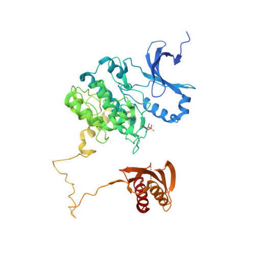
4CFF | pdb_00004cff
Structure of full length human AMPK in complex with a small molecule activator, a thienopyridone derivative (A-769662)
- PDB DOI: https://doi.org/10.2210/pdb4CFF/pdb
- Classification: TRANSFERASE
- Organism(s): Homo sapiens
- Expression System: Escherichia coli
- Mutation(s): No
- Deposited: 2013-11-14 Released: 2013-12-25
- Deposition Author(s): Xiao, B., Sanders, M.J., Carmena, D., Bright, N.J., Haire, L.F., Underwood, E., Patel, B.R., Heath, R.B., Walker, P.A., Hallen, S., Giordanetto, F., Martin, S.R., Carling, D., Gamblin, S.J.
Experimental Data Snapshot
- Method: X-RAY DIFFRACTION
- Resolution: 3.92 Å
- R-Value Free: 0.264 (Depositor), 0.260 (DCC)
- R-Value Work: 0.208 (Depositor), 0.210 (DCC)
- R-Value Observed: 0.211 (Depositor)
Starting Models: experimental
View more details
- 👁 Image
Download Mendeley
Structural Basis of Ampk Regulation by Small Molecule Activators.
Xiao, B., Sanders, M.J., Carmena, D., Bright, N.J., Haire, L.F., Underwood, E., Patel, B.R., Heath, R.B., Walker, P.A., Hallen, S., Giordanetto, F., Martin, S.R., Carling, D., Gamblin, S.J.(2013) Nat Commun 4: 3017
- PubMed: 24352254 Search on PubMedSearch on PubMed Central
- DOI: https://doi.org/10.1038/ncomms4017
- Primary Citation Related Structures:
4CFE, 4CFF - PubMed Abstract:
AMP-activated protein kinase (AMPK) plays a major role in regulating cellular energy balance by sensing and responding to increases in AMP/ADP concentration relative to ATP. Binding of AMP causes allosteric activation of the enzyme and binding of either AMP or ADP promotes and maintains the phosphorylation of threonine 172 within the activation loop of the kinase. AMPK has attracted widespread interest as a potential therapeutic target for metabolic diseases including type 2 diabetes and, more recently, cancer. A number of direct AMPK activators have been reported as having beneficial effects in treating metabolic diseases, but there has been no structural basis for activator binding to AMPK. Here we present the crystal structure of human AMPK in complex with a small molecule activator that binds at a site between the kinase domain and the carbohydrate-binding module, stabilising the interaction between these two components. The nature of the activator-binding pocket suggests the involvement of an additional, as yet unidentified, metabolite in the physiological regulation of AMPK. Importantly, the structure offers new opportunities for the design of small molecule activators of AMPK for treatment of metabolic disorders.
- 1] MRC National Institute for Medical Research, The Ridgeway, Mill Hill, London NW7 1AA, UK [2].
- 1] MRC Clinical Sciences Centre, Cellular Stress Group, Hammersmith Hospital Campus, Imperial College, DuCane Road, London W12 0NN, UK [2].
- MRC Clinical Sciences Centre, Cellular Stress Group, Hammersmith Hospital Campus, Imperial College, DuCane Road, London W12 0NN, UK.
- MRC National Institute for Medical Research, The Ridgeway, Mill Hill, London NW7 1AA, UK.
- Bioscience, CVMD Innovative Medicine Unit, AstraZeneca R&D, Pepparedsleden 1, Mölndal S-43183, Sweden.
- 1] Medicinal Chemistry, CVMD Innovative Medicine Unit, AstraZeneca R&D, Pepparedsleden 1, Mölndal S-43183, Sweden [2].
Organizational Affiliation:
Explore in 3D: Structure | Sequence Annotations | Electron Density | Validation Report | Ligand Interaction (STU)
Explore in 3D: Structure | Sequence Annotations | Electron Density | Validation Report | Ligand Interaction (STU)
Global Symmetry: Asymmetric - C1
Global Stoichiometry: Hetero 3-mer - A1B1C1
Find Similar Assemblies
Biological assembly 1 assigned by authors and generated by PISA (software)
Explore in 3D: Structure | Sequence Annotations | Electron Density | Validation Report | Ligand Interaction (STU)
Global Symmetry: Asymmetric - C1
Global Stoichiometry: Hetero 3-mer - A1B1C1
Find Similar Assemblies
Biological assembly 2 assigned by authors and generated by PISA (software)
Macromolecule Content
- Total Structure Weight: 272.49 kDa
- Atom Count: 14,484
- Modeled Residue Count: 1,782
- Deposited Residue Count: 2,376
- Unique protein chains: 3
Entity ID: 1 | |||||
|---|---|---|---|---|---|
| Molecule | Chains | Sequence Length | Organism | Details | Image |
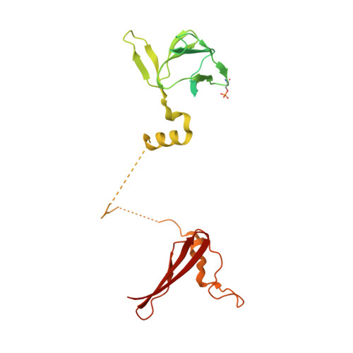
| 5'-AMP-ACTIVATED PROTEIN KINASE CATALYTIC SUBUNIT ALPHA-2 | 571 | Homo sapiens | Mutation(s): 0 EC: 2.7.11.1 (PDB Primary Data), 2.7.11.27 (PDB Primary Data), 2.7.11.31 (PDB Primary Data) | 👁 Image | |
UniProt & NIH Common Fund Data Resources | |||||
Find proteins for P54646 (Homo sapiens) Explore P54646 Go to UniProtKB: P54646 | |||||
PHAROS: P54646 GTEx: ENSG00000162409 | |||||
Entity Groups | |||||
| Sequence Clusters | 30% Identity50% Identity70% Identity90% Identity95% Identity100% Identity | ||||
| UniProt Group | P54646 | ||||
Sequence AnnotationsExpand | |||||
| |||||
Entity ID: 2 | |||||
|---|---|---|---|---|---|
| Molecule | Chains | Sequence Length | Organism | Details | Image |
| 5'-AMP-ACTIVATED PROTEIN KINASE SUBUNIT BETA-1 | 286 | Homo sapiens | Mutation(s): 0 | 👁 Image | |
UniProt & NIH Common Fund Data Resources | |||||
Find proteins for Q9Y478 (Homo sapiens) Explore Q9Y478 Go to UniProtKB: Q9Y478 | |||||
PHAROS: Q9Y478 GTEx: ENSG00000111725 | |||||
Entity Groups | |||||
| Sequence Clusters | 30% Identity50% Identity70% Identity90% Identity95% Identity100% Identity | ||||
| UniProt Group | Q9Y478 | ||||
Sequence AnnotationsExpand | |||||
| |||||
Entity ID: 3 | |||||
|---|---|---|---|---|---|
| Molecule | Chains | Sequence Length | Organism | Details | Image |
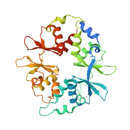
| 5'-AMP-ACTIVATED PROTEIN KINASE SUBUNIT GAMMA-1 | 331 | Homo sapiens | Mutation(s): 0 | 👁 Image | |
UniProt & NIH Common Fund Data Resources | |||||
Find proteins for P54619 (Homo sapiens) Explore P54619 Go to UniProtKB: P54619 | |||||
PHAROS: P54619 GTEx: ENSG00000181929 | |||||
Entity Groups | |||||
| Sequence Clusters | 30% Identity50% Identity70% Identity90% Identity95% Identity100% Identity | ||||
| UniProt Group | P54619 | ||||
Sequence AnnotationsExpand | |||||
| |||||
| Ligands 3 Unique | |||||
|---|---|---|---|---|---|
| ID | Chains | Name / Formula / InChI Key | 2D Diagram | 3D Interactions | |
| STU Query on STU Download Ideal Coordinates CCD File | G [auth A], I [auth C] | STAUROSPORINE C28 H26 N4 O3 HKSZLNNOFSGOKW-FYTWVXJKSA-N | 👁 Image | ||
| C1V Query on C1V Download Ideal Coordinates CCD File | H [auth B], J [auth C] | 3-[4-(2-hydroxyphenyl)phenyl]-4-oxidanyl-6-oxidanylidene-7H-thieno[2,3-b]pyridine-5-carbonitrile C20 H12 N2 O3 S CTESJDQKVOEUOY-UHFFFAOYSA-N | 👁 Image | ||
| AMP Query on AMP Download Ideal Coordinates CCD File
| K [auth E], L [auth E], M [auth F], N [auth F] | ADENOSINE MONOPHOSPHATE C10 H14 N5 O7 P UDMBCSSLTHHNCD-KQYNXXCUSA-N | 👁 Image | ||
| Modified Residues 2 Unique | |||||
|---|---|---|---|---|---|
| ID | Chains | Type | Formula | 2D Diagram | Parent |
| TPO Query on TPO | A, C | L-PEPTIDE LINKING | C4 H10 N O6 P | 👁 Image | THR |
| SEP Query on SEP | B, D | L-PEPTIDE LINKING | C3 H8 N O6 P | 👁 Image | SER |
Experimental Data
- Method: X-RAY DIFFRACTION
- Resolution: 3.92 Å
- R-Value Free: 0.264 (Depositor), 0.260 (DCC)
- R-Value Work: 0.208 (Depositor), 0.210 (DCC)
- R-Value Observed: 0.211 (Depositor)
| Length ( Å ) | Angle ( ˚ ) |
|---|---|
| a = 76.019 | α = 90 |
| b = 134.787 | β = 93.04 |
| c = 141.286 | γ = 90 |
| Software Name | Purpose |
|---|---|
| PHENIX | refinement |
| DENZO | data reduction |
| SCALEPACK | data scaling |
| PHASER | phasing |
Deposition Data
- Released Date: 2013-12-25 Deposition Author(s): Xiao, B., Sanders, M.J., Carmena, D., Bright, N.J., Haire, L.F., Underwood, E., Patel, B.R., Heath, R.B., Walker, P.A., Hallen, S., Giordanetto, F., Martin, S.R., Carling, D., Gamblin, S.J.
Revision History (Full details and data files)
- Version 1.0: 2013-12-25
Type: Initial release - Version 1.1: 2014-01-08
Changes: Database references - Version 1.2: 2014-02-12
Changes: Data collection, Other, Refinement description - Version 1.3: 2019-05-08
Changes: Data collection, Derived calculations, Experimental preparation, Other - Version 1.4: 2023-12-20
Changes: Data collection, Database references, Derived calculations, Other, Refinement description - Version 1.5: 2024-10-23
Changes: Structure summary