
- FASTA Sequence
- PDBx/mmCIF Format
- PDBx/mmCIF Format (gz)
- BinaryCIF Format (gz)
- Legacy PDB Format
- Legacy PDB Format (gz)
- PDBML/XML Format (gz)
- Structure Factors (CIF)
- Structure Factors (CIF - gz)
- Validation Full (PDF - gz)
- Validation (XML - gz)
- Validation (CIF - gz)
- Validation 2fo-fc coefficients (CIF - gz)
- Validation fo-fc coefficients (CIF - gz)
- Biological Assembly 1 (CIF - gz)
- Biological Assembly 1 (PDB - gz)
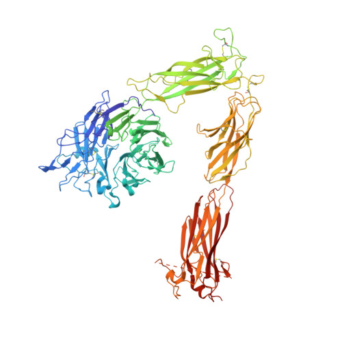
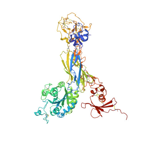
4G1M | pdb_00004g1m
Re-refinement of alpha V beta 3 structure
- PDB DOI: https://doi.org/10.2210/pdb4G1M/pdb
- Classification: PROTEIN BINDING
- Organism(s): Homo sapiens
- Expression System: Cricetulus griseus
- Mutation(s): No
- Deposited: 2012-07-10 Released: 2012-12-12
- Deposition Author(s): Springer, T.A., Mi, L., Zhu, J.
Experimental Data Snapshot
- Method: X-RAY DIFFRACTION
- Resolution: 2.90 Å
- R-Value Free: 0.233 (Depositor), 0.241 (DCC)
- R-Value Work: 0.179 (Depositor), 0.186 (DCC)
- R-Value Observed: 0.182 (Depositor)
Literature
- 👁 Image
Download Mendeley
Alpha V Beta 3 Integrin Crystal Structures and their Functional Implications
Dong, X., Mi, L.Z., Zhu, J., Wang, W., Hu, P., Luo, B.H., Springer, T.A.(2012) Biochemistry 51: 8814-8828
- PubMed: 23106217 Search on PubMedSearch on PubMed Central
- DOI: https://doi.org/10.1021/bi300734n
- Primary Citation Related Structures:
4G1E, 4G1M - PubMed Abstract:
Many questions about the significance of structural features of integrin α(V)β(3) with respect to its mechanism of activation remain. We have determined and re-refined crystal structures of the α(V)β(3) ectodomain linked to C-terminal coiled coils (α(V)β(3)-AB) and four transmembrane (TM) residues in each subunit (α(V)β(3)-1TM), respectively. The α(V) and β(3) subunits with four and eight extracellular domains, respectively, are bent at knees between the integrin headpiece and lower legs, and the headpiece has the closed, low-affinity conformation. The structures differ in the occupancy of three metal-binding sites in the βI domain. Occupancy appears to be related to the pH of crystallization, rather than to the physiologic regulation of ligand binding at the central, metal ion-dependent adhesion site. No electron density was observed for TM residues and much of the α(V) linker. α(V)β(3)-AB and α(V)β(3)-1TM demonstrate flexibility in the linker between their extracellular and TM domains, rather than the previously proposed rigid linkage. A previously postulated interface between the α(V) and β(3) subunits at their knees was also not supported, because it lacks high-quality density, required rebuilding in α(V)β(3)-1TM, and differed markedly between α(V)β(3)-1TM and α(V)β(3)-AB. Together with the variation in domain-domain orientation within their bent ectodomains between α(V)β(3)-AB and α(V)β(3)-1TM, these findings are compatible with the requirement for large structural changes, such as extension at the knees and headpiece opening, in conveying activation signals between the extracellular ligand-binding site and the cytoplasm.
- Immune Disease Institute, Children's Hospital Boston, and Department of Biological Chemistry and Molecular Pharmacology, Harvard Medical School, 3 Blackfan Circle, Boston, Massachusetts 02115, United States.
Organizational Affiliation:
Explore in 3D: Structure | Sequence Annotations | Electron Density | Validation Report | Ligand Interaction (NAG)
Biological Assembly 1
Explore in 3D: Structure | Sequence Annotations | Electron Density | Validation Report | Ligand Interaction (NAG)
Global Symmetry: Asymmetric - C1
Global Stoichiometry: Hetero 2-mer - A1B1
Find Similar Assemblies
Biological assembly 1 assigned by authors and generated by PISA (software)
Macromolecule Content
- Total Structure Weight: 190.31 kDa
- Atom Count: 13,132
- Modeled Residue Count: 1,621
- Deposited Residue Count: 1,651
- Unique protein chains: 2
Macromolecules
Entity ID: 1 | |||||
|---|---|---|---|---|---|
| Molecule | Chains | Sequence Length | Organism | Details | Image |
| Integrin alpha-V | 959 | Homo sapiens | Mutation(s): 0 Gene Names: ITGAV, MSK8, VNRA | 👁 Image | |
UniProt & NIH Common Fund Data Resources | |||||
PHAROS: P06756 GTEx: ENSG00000138448 | |||||
Entity Groups | |||||
| Sequence Clusters | 30% Identity50% Identity70% Identity90% Identity95% Identity100% Identity | ||||
| UniProt Group | P06756 | ||||
Glycosylation | |||||
| Glycosylation Sites: 11 | Go to GlyGen: P06756-1 | ||||
Sequence AnnotationsExpand | |||||
Reference Sequence | |||||
Entity ID: 2 | |||||
|---|---|---|---|---|---|
| Molecule | Chains | Sequence Length | Organism | Details | Image |
| Integrin beta-3 | 692 | Homo sapiens | Mutation(s): 0 Gene Names: ITGB3, GP3A | 👁 Image | |
UniProt & NIH Common Fund Data Resources | |||||
PHAROS: P05106 GTEx: ENSG00000259207 | |||||
Entity Groups | |||||
| Sequence Clusters | 30% Identity50% Identity70% Identity90% Identity95% Identity100% Identity | ||||
| UniProt Group | P05106 | ||||
Glycosylation | |||||
| Glycosylation Sites: 5 | Go to GlyGen: P05106-1 | ||||
Sequence AnnotationsExpand | |||||
Reference Sequence | |||||
Oligosaccharides
HelpEntity ID: 3 | |||||
|---|---|---|---|---|---|
| Molecule | Chains | Length | 2D Diagram | Glycosylation | D Interactions |
| alpha-D-mannopyranose-(1-3)-[alpha-D-mannopyranose-(1-6)]beta-D-mannopyranose-(1-4)-2-acetamido-2-deoxy-beta-D-glucopyranose-(1-4)-2-acetamido-2-deoxy-beta-D-glucopyranose | C | 5 | 👁 Image | N-Glycosylation | |
Glycosylation Resources | |||||
GlyTouCan: G22768VO GlyCosmos: G22768VO GlyGen: G22768VO | |||||
Entity ID: 4 | |||||
|---|---|---|---|---|---|
| Molecule | Chains | Length | 2D Diagram | Glycosylation | D Interactions |
| 2-acetamido-2-deoxy-beta-D-glucopyranose-(1-4)-2-acetamido-2-deoxy-beta-D-glucopyranose | D, F, G, H, I D, F, G, H, I, J, K, L, M | 2 | 👁 Image | N-Glycosylation | |
Glycosylation Resources | |||||
GlyTouCan: G42666HT GlyCosmos: G42666HT GlyGen: G42666HT | |||||
Entity ID: 5 | |||||
|---|---|---|---|---|---|
| Molecule | Chains | Length | 2D Diagram | Glycosylation | D Interactions |
| alpha-D-mannopyranose-(1-2)-alpha-D-mannopyranose-(1-3)-[alpha-D-mannopyranose-(1-3)-alpha-D-mannopyranose-(1-6)]beta-D-mannopyranose-(1-4)-2-acetamido-2-deoxy-beta-D-glucopyranose-(1-4)-2-acetamido-2-deoxy-beta-D-glucopyranose | E | 7 | 👁 Image | N-Glycosylation | |
Glycosylation Resources | |||||
GlyTouCan: G07617FP GlyCosmos: G07617FP GlyGen: G07617FP | |||||
Small Molecules
Experimental Data & Validation
Experimental Data
- Method: X-RAY DIFFRACTION
- Resolution: 2.90 Å
- R-Value Free: 0.233 (Depositor), 0.241 (DCC)
- R-Value Work: 0.179 (Depositor), 0.186 (DCC)
- R-Value Observed: 0.182 (Depositor)
| Length ( Å ) | Angle ( ˚ ) |
|---|---|
| a = 129.87 | α = 90 |
| b = 129.87 | β = 90 |
| c = 305.9 | γ = 120 |
| Software Name | Purpose |
|---|---|
| PHENIX | refinement |
| CNS | refinement |
| HKL-2000 | data collection |
| DENZO | data reduction |
| HKL-2000 | data scaling |
| CNS | phasing |
Entry History
Deposition Data
- Released Date: 2012-12-12 Deposition Author(s): Springer, T.A., Mi, L., Zhu, J.
Revision History (Full details and data files)
- Version 1.0: 2012-12-12
Type: Initial release - Version 1.1: 2012-12-19
Changes: Database references - Version 1.2: 2019-08-14
Changes: Data collection, Derived calculations, Refinement description - Version 2.0: 2020-07-29
Type: Remediation
Reason: Carbohydrate remediation
Changes: Advisory, Atomic model, Data collection, Derived calculations, Structure summary - Version 2.1: 2024-11-06
Changes: Data collection, Database references, Derived calculations, Structure summary