
- FASTA Sequence
- PDBx/mmCIF Format
- PDBx/mmCIF Format (gz)
- BinaryCIF Format (gz)
- Legacy PDB Format
- Legacy PDB Format (gz)
- PDBML/XML Format (gz)
- Structure Factors (CIF)
- Structure Factors (CIF - gz)
- Validation Full (PDF - gz)
- Validation (XML - gz)
- Validation (CIF - gz)
- Validation 2fo-fc coefficients (CIF - gz)
- Validation fo-fc coefficients (CIF - gz)
- Biological Assembly 1 (CIF - gz)
- Biological Assembly 1 (PDB - gz)
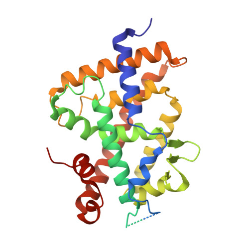
4IA3 | pdb_00004ia3
Diastereotopic and Deuterium Effects in Gemini
- PDB DOI: https://doi.org/10.2210/pdb4IA3/pdb
- Classification: TRANSCRIPTION/TRANSCRIPTION ACTIVATOR
- Organism(s): Danio rerio, Homo sapiens
- Expression System: Escherichia coli
- Mutation(s): No
- Deposited: 2012-12-06 Released: 2013-04-24
- Deposition Author(s): Maehr, H., Rochel, N., Suh, N., Uskokovic, M.
Experimental Data Snapshot
- Method: X-RAY DIFFRACTION
- Resolution: 2.70 Å
- R-Value Free: 0.260 (Depositor), 0.250 (DCC)
- R-Value Work: 0.209 (Depositor), 0.210 (DCC)
- R-Value Observed: 0.209 (Depositor)
- 👁 Image
Download Mendeley
Diastereotopic and deuterium effects in gemini.
Maehr, H., Rochel, N., Lee, H.J., Suh, N., Uskokovic, M.R.(2013) J Med Chem 56: 3878-3888
- PubMed: 23566225 Search on PubMed
- DOI: https://doi.org/10.1021/jm400032t
- Primary Citation Related Structures:
4IA1, 4IA2, 4IA3, 4IA7 - PubMed Abstract:
Changing the geminal methyl groups on 1α,25-dihydroxyvitamin D3 and its analogues to the deuterio versions generally improves the bioactivity. Derivatives of 1α,25-dihydroxyvitamin D3 with two chains emanating at C20, commonly referred to as gemini, are subject to the same phenomenon. Additionally, gemini with different side chains are susceptible to bioactivity differentials where the C17-C20 threo configuration usually imparts higher activity than the corresponding erythro arrangement. In an effort to analyze the deuterium effect on gemini with minimal diastereotopic distortion, we synthesized gemini with equal side chains but introduced deuterium diastereospecifically on either chain. We solved the crystal structures of these compounds in the zebra fish zVDR ligand binding domain as complexes with NCoA-2 coactivator peptide and correlated the findings with growth inhibition in a breast cancer cell line.
- Department of Chemical Biology, Ernest Mario School of Pharmacy, Rutgers, The State University of New Jersey, 164 Frelinghuysen Road, Piscataway, New Jersey 08854, USA. hubert.maehr@verizon.net
Organizational Affiliation:
Explore in 3D: Structure | Sequence Annotations | Electron Density | Validation Report | Ligand Interaction (BIV)
Explore in 3D: Structure | Sequence Annotations | Electron Density | Validation Report | Ligand Interaction (BIV)
Global Symmetry: Cyclic - C2 (Explore in 3D)
Global Stoichiometry: Hetero 4-mer - A2B2
Pseudo Symmetry: Asymmetric - C1
Pseudo Stoichiometry: Hetero 4-mer - A2B1C1
Find Similar Assemblies
Biological assembly 1 assigned by authors and generated by PISA (software)
Macromolecule Content
- Total Structure Weight: 36 kDa
- Atom Count: 2,111
- Modeled Residue Count: 249
- Deposited Residue Count: 313
- Unique protein chains: 2
Entity ID: 1 | |||||
|---|---|---|---|---|---|
| Molecule | Chains | Sequence Length | Organism | Details | Image |
| Vitamin D3 receptor A | 300 | Danio rerio | Mutation(s): 0 Gene Names: vdra, nr1i1a, vdr | 👁 Image | |
UniProt | |||||
Find proteins for Q9PTN2 (Danio rerio) Explore Q9PTN2 Go to UniProtKB: Q9PTN2 | |||||
Entity Groups | |||||
| Sequence Clusters | 30% Identity50% Identity70% Identity90% Identity95% Identity100% Identity | ||||
| UniProt Group | Q9PTN2 | ||||
Sequence AnnotationsExpand | |||||
| |||||
Find similar proteins by: Sequence | 3D Structure
Entity ID: 2 | |||||
|---|---|---|---|---|---|
| Molecule | Chains | Sequence Length | Organism | Details | Image |
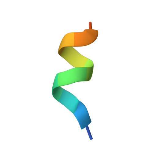
| Nuclear receptor coactivator 2 | 13 | Homo sapiens | Mutation(s): 0 | 👁 Image | |
UniProt & NIH Common Fund Data Resources | |||||
Find proteins for Q15596 (Homo sapiens) Explore Q15596 Go to UniProtKB: Q15596 | |||||
PHAROS: Q15596 GTEx: ENSG00000140396 | |||||
Entity Groups | |||||
| Sequence Clusters | 30% Identity50% Identity70% Identity90% Identity95% Identity100% Identity | ||||
| UniProt Group | Q15596 | ||||
Sequence AnnotationsExpand | |||||
| |||||
| Ligands 1 Unique | |||||
|---|---|---|---|---|---|
| ID | Chains | Name / Formula / InChI Key | 2D Diagram | 3D Interactions | |
| BIV Query on BIV Download Ideal Coordinates CCD File | C [auth A] | 21-NOR-9,10-SECOCHOLESTA-5,7,10(19)-TRIENE-1,3,25-TRIOL, 20-(4-HYDROXY-4-METHYLPENTYL)-, (1A,3B,5Z,7E) C32 H54 O4 WTQXZYVWLNPNEX-LKUPKUSPSA-N | 👁 Image | ||
Experimental Data
- Method: X-RAY DIFFRACTION
- Resolution: 2.70 Å
- R-Value Free: 0.260 (Depositor), 0.250 (DCC)
- R-Value Work: 0.209 (Depositor), 0.210 (DCC)
- R-Value Observed: 0.209 (Depositor)
| Length ( Å ) | Angle ( ˚ ) |
|---|---|
| a = 66.044 | α = 90 |
| b = 66.044 | β = 90 |
| c = 266.634 | γ = 120 |
| Software Name | Purpose |
|---|---|
| CNS | refinement |
| HKL-2000 | data reduction |
| HKL-2000 | data scaling |
| CNS | phasing |
Deposition Data
- Released Date: 2013-04-24 Deposition Author(s): Maehr, H., Rochel, N., Suh, N., Uskokovic, M.
Revision History (Full details and data files)
- Version 1.0: 2013-04-24
Type: Initial release - Version 1.1: 2013-07-10
Changes: Database references - Version 1.2: 2024-02-28
Changes: Data collection, Database references, Derived calculations