
- FASTA Sequence
- PDBx/mmCIF Format
- PDBx/mmCIF Format (gz)
- BinaryCIF Format (gz)
- Legacy PDB Format
- Legacy PDB Format (gz)
- PDBML/XML Format (gz)
- Structure Factors (CIF)
- Structure Factors (CIF - gz)
- Validation Full (PDF - gz)
- Validation (XML - gz)
- Validation (CIF - gz)
- Validation 2fo-fc coefficients (CIF - gz)
- Validation fo-fc coefficients (CIF - gz)
- Biological Assembly 1 (CIF - gz)
- Biological Assembly 1 (PDB - gz)
4MMZ | pdb_00004mmz
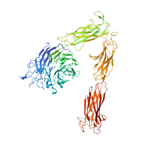
Integrin AlphaVBeta3 ectodomain bound to an antagonistic tenth domain of Fibronectin
- PDB DOI: https://doi.org/10.2210/pdb4MMZ/pdb
- Classification: CELL ADHESION
- Organism(s): Homo sapiens
- Expression System: Spodoptera frugiperda, Escherichia coli BL21
- Mutation(s): Yes
- Deposited: 2013-09-09 Released: 2014-03-26
- Deposition Author(s): van Agthoven, J., Xiong, J., Arnaout, M.A.
- Funding Organization(s): National Institutes of Health/National Institute of Diabetes and Digestive and Kidney Disease (NIH/NIDDK)
Experimental Data Snapshot
- Method: X-RAY DIFFRACTION
- Resolution: 3.10 Å
- R-Value Free: 0.255 (Depositor), 0.255 (DCC)
- R-Value Work: 0.207 (Depositor), 0.207 (DCC)
- R-Value Observed: 0.209 (Depositor)
Starting Model: experimental
View more details
Literature
- 👁 Image
Download Mendeley
Structural basis for pure antagonism of integrin alpha V beta 3 by a high-affinity form of fibronectin.
Van Agthoven, J.F., Xiong, J.P., Alonso, J.L., Rui, X., Adair, B.D., Goodman, S.L., Arnaout, M.A.(2014) Nat Struct Mol Biol 21: 383-388
- PubMed: 24658351 Search on PubMedSearch on PubMed Central
- DOI: https://doi.org/10.1038/nsmb.2797
- Primary Citation Related Structures:
4MMX, 4MMY, 4MMZ - PubMed Abstract:
Integrins are important therapeutic targets. However, current RGD-based anti-integrin drugs are also partial agonists, inducing conformational changes that trigger potentially fatal immune reactions and paradoxical cell adhesion. Here we describe the first crystal structure of αVβ3 bound to a physiologic ligand, the tenth type III RGD domain of wild-type fibronectin (wtFN10), or to a high-affinity mutant (hFN10) shown here to act as a pure antagonist. Comparison of these structures revealed a central π-π interaction between Trp1496 in the RGD-containing loop of hFN10 and Tyr122 of the β3 subunit that blocked conformational changes triggered by wtFN10 and trapped hFN10-bound αVβ3 in an inactive conformation. Removing the Trp1496 or Tyr122 side chains or reorienting Trp1496 away from Tyr122 converted hFN10 into a partial agonist. These findings offer new insights into the mechanism of integrin activation and a basis for the design of RGD-based pure antagonists.
- 1] Structural Biology Program, Department of Medicine, Harvard Medical School, Massachusetts General Hospital, Charlestown, Massachusetts, USA. [2].
- Leukocyte Biology and Inflammation Program, Department of Medicine, Harvard Medical School, Massachusetts General Hospital, Charlestown, Massachusetts, USA.
- Structural Biology Program, Department of Medicine, Harvard Medical School, Massachusetts General Hospital, Charlestown, Massachusetts, USA.
- Global Research and Early Development, Translational Innovation Platform, Oncology, Merck KGaA, Darmstadt, Germany.
- 1] Structural Biology Program, Department of Medicine, Harvard Medical School, Massachusetts General Hospital, Charlestown, Massachusetts, USA. [2] Leukocyte Biology and Inflammation Program, Department of Medicine, Harvard Medical School, Massachusetts General Hospital, Charlestown, Massachusetts, USA.
Organizational Affiliation:
Explore in 3D: Structure | Sequence Annotations | Electron Density | Validation Report | Ligand Interaction (NAG)
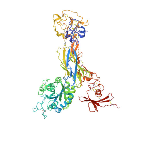
Biological Assembly 1
Explore in 3D: Structure | Sequence Annotations | Electron Density | Validation Report | Ligand Interaction (NAG)
Global Symmetry: Asymmetric - C1
Global Stoichiometry: Hetero 3-mer - A1B1C1
Find Similar Assemblies
Biological assembly 1 assigned by authors.
Macromolecule Content
- Total Structure Weight: 198.88 kDa
- Atom Count: 13,512
- Modeled Residue Count: 1,701
- Deposited Residue Count: 1,749
- Unique protein chains: 3
Macromolecules
Entity ID: 1 | |||||
|---|---|---|---|---|---|
| Molecule | Chains | Sequence Length | Organism | Details | Image |
| Integrin alpha-V | 959 | Homo sapiens | Mutation(s): 0 Gene Names: ITGAV, MSK8, VNRA, VTNR | 👁 Image | |
UniProt & NIH Common Fund Data Resources | |||||
PHAROS: P06756 GTEx: ENSG00000138448 | |||||
Entity Groups | |||||
| Sequence Clusters | 30% Identity50% Identity70% Identity90% Identity95% Identity100% Identity | ||||
| UniProt Group | P06756 | ||||
Glycosylation | |||||
| Glycosylation Sites: 9 | Go to GlyGen: P06756-1 | ||||
Sequence AnnotationsExpand | |||||
Reference Sequence | |||||
Entity ID: 2 | |||||
|---|---|---|---|---|---|
| Molecule | Chains | Sequence Length | Organism | Details | Image |
| Integrin beta-3 | 692 | Homo sapiens | Mutation(s): 0 Gene Names: ITGB3, GP3A | 👁 Image | |
UniProt & NIH Common Fund Data Resources | |||||
PHAROS: P05106 GTEx: ENSG00000259207 | |||||
Entity Groups | |||||
| Sequence Clusters | 30% Identity50% Identity70% Identity90% Identity95% Identity100% Identity | ||||
| UniProt Group | P05106 | ||||
Glycosylation | |||||
| Glycosylation Sites: 5 | Go to GlyGen: P05106-1 | ||||
Sequence AnnotationsExpand | |||||
Reference Sequence | |||||
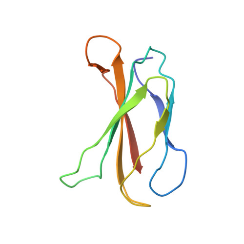
Entity ID: 3 | |||||
|---|---|---|---|---|---|
| Molecule | Chains | Sequence Length | Organism | Details | Image |
| Fibronectin | 98 | Homo sapiens | Mutation(s): 5 Gene Names: FN1, FN | 👁 Image | |
UniProt & NIH Common Fund Data Resources | |||||
PHAROS: P02751 GTEx: ENSG00000115414 | |||||
Entity Groups | |||||
| Sequence Clusters | 30% Identity50% Identity70% Identity90% Identity95% Identity100% Identity | ||||
| UniProt Group | P02751 | ||||
Sequence AnnotationsExpand | |||||
Reference Sequence | |||||
Oligosaccharides
HelpEntity ID: 4 | |||||
|---|---|---|---|---|---|
| Molecule | Chains | Length | 2D Diagram | Glycosylation | D Interactions |
| 2-acetamido-2-deoxy-beta-D-glucopyranose-(1-4)-2-acetamido-2-deoxy-beta-D-glucopyranose | D, F, G, H | 2 | 👁 Image | N-Glycosylation | |
Glycosylation Resources | |||||
GlyTouCan: G42666HT GlyCosmos: G42666HT GlyGen: G42666HT | |||||
Entity ID: 5 | |||||
|---|---|---|---|---|---|
| Molecule | Chains | Length | 2D Diagram | Glycosylation | D Interactions |
| alpha-D-mannopyranose-(1-4)-beta-D-mannopyranose-(1-6)-[alpha-D-mannopyranose-(1-3)]beta-D-mannopyranose-(1-4)-2-acetamido-2-deoxy-beta-D-glucopyranose-(1-4)-2-acetamido-2-deoxy-beta-D-glucopyranose | E | 6 | 👁 Image | N-Glycosylation | |
Glycosylation Resources | |||||
GlyTouCan: G93290PO GlyCosmos: G93290PO GlyGen: G93290PO | |||||
Small Molecules
Experimental Data & Validation
Experimental Data
- Method: X-RAY DIFFRACTION
- Resolution: 3.10 Å
- R-Value Free: 0.255 (Depositor), 0.255 (DCC)
- R-Value Work: 0.207 (Depositor), 0.207 (DCC)
- R-Value Observed: 0.209 (Depositor)
| Length ( Å ) | Angle ( ˚ ) |
|---|---|
| a = 129.792 | α = 90 |
| b = 129.792 | β = 90 |
| c = 307.675 | γ = 120 |
| Software Name | Purpose |
|---|---|
| DENZO | data reduction |
| SCALEPACK | data scaling |
| PHASER | phasing |
| PHENIX | refinement |
| HKL-2000 | data reduction |
| HKL-2000 | data scaling |
Entry History
& Funding InformationDeposition Data
- Released Date: 2014-03-26 Deposition Author(s): van Agthoven, J., Xiong, J., Arnaout, M.A.
| Funding Organization | Location | Grant Number |
|---|---|---|
| National Institutes of Health/National Institute of Diabetes and Digestive and Kidney Disease (NIH/NIDDK) | United States | -- |
Revision History (Full details and data files)
- Version 1.0: 2014-03-26
Type: Initial release - Version 1.1: 2014-04-09
Changes: Database references - Version 1.2: 2014-04-30
Changes: Database references - Version 1.3: 2017-11-15
Changes: Refinement description - Version 2.0: 2020-07-29
Type: Remediation
Reason: Carbohydrate remediation
Changes: Atomic model, Data collection, Database references, Derived calculations, Structure summary - Version 3.0: 2022-12-21
Type: Coordinate replacement
Reason: Atomic clashes
Changes: Atomic model, Author supporting evidence, Data collection, Database references, Derived calculations, Non-polymer description, Other, Refinement description, Source and taxonomy, Structure summary - Version 3.1: 2023-09-20
Changes: Data collection, Refinement description - Version 3.2: 2024-11-20
Changes: Structure summary