
- FASTA Sequence
- PDBx/mmCIF Format
- PDBx/mmCIF Format (gz)
- BinaryCIF Format (gz)
- Legacy PDB Format
- Legacy PDB Format (gz)
- PDBML/XML Format (gz)
- Structure Factors (CIF)
- Structure Factors (CIF - gz)
- Validation Full (PDF - gz)
- Validation (XML - gz)
- Validation (CIF - gz)
- Validation 2fo-fc coefficients (CIF - gz)
- Validation fo-fc coefficients (CIF - gz)
- Biological Assembly 1 (CIF - gz)
- Biological Assembly 2 (CIF - gz)
- Biological Assembly 3 (CIF - gz)
- Biological Assembly 4 (CIF - gz)
- Biological Assembly 5 (CIF - gz)
- Biological Assembly 6 (CIF - gz)
- Biological Assembly 1 (PDB - gz)
- Biological Assembly 2 (PDB - gz)
- Biological Assembly 3 (PDB - gz)
- Biological Assembly 4 (PDB - gz)
- Biological Assembly 5 (PDB - gz)
- Biological Assembly 6 (PDB - gz)
4N34 | pdb_00004n34
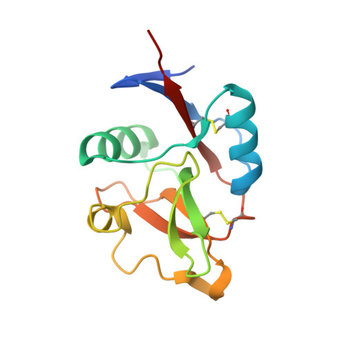
Structure of langerin CRD I313 with alpha-MeGlcNAc
- PDB DOI: https://doi.org/10.2210/pdb4N34/pdb
- Classification: SUGAR BINDING PROTEIN
- Organism(s): Homo sapiens
- Expression System: Escherichia coli
- Mutation(s): Yes
- Deposited: 2013-10-06 Released: 2013-11-20
- Deposition Author(s): Feinberg, H., Rowntree, T.J.W., Tan, S.L.W., Drickamer, K., Weis, W.I., Taylor, M.E.
Experimental Data Snapshot
- Method: X-RAY DIFFRACTION
- Resolution: 1.75 Å
- R-Value Free: 0.229 (Depositor), 0.249 (DCC)
- R-Value Work: 0.176 (Depositor)
- R-Value Observed: 0.178 (Depositor)
Starting Model: experimental
View more details
Literature
- 👁 Image
Download Mendeley
Common polymorphisms in human langerin change specificity for glycan ligands.
Feinberg, H., Rowntree, T.J., Tan, S.L., Drickamer, K., Weis, W.I., Taylor, M.E.(2013) J Biological Chem 288: 36762-36771
- PubMed: 24217250 Search on PubMedSearch on PubMed Central
- DOI: https://doi.org/10.1074/jbc.M113.528000
- Primary Citation Related Structures:
4N32, 4N33, 4N34, 4N35, 4N36, 4N37, 4N38 - PubMed Abstract:
Langerin, a C-type lectin on Langerhans cells, mediates carbohydrate-dependent uptake of pathogens in the first step of antigen presentation to the adaptive immune system. Langerin binds a diverse range of carbohydrates including high mannose structures, fucosylated blood group antigens, and glycans with terminal 6-sulfated galactose. Mutagenesis and quantitative binding assays indicate that salt bridges between the sulfate group and two lysine residues compensate for the nonoptimal binding of galactose at the primary Ca(2+) site. A commonly occurring single nucleotide polymorphism (SNP) in human langerin results in change of one of these lysine residues, Lys-313, to isoleucine. Glycan array screening reveals that this amino acid change abolishes binding to oligosaccharides with terminal 6SO4-Gal and enhances binding to oligosaccharides with terminal GlcNAc residues. Structural analysis shows that enhanced binding to GlcNAc may result from Ile-313 packing against the N-acetyl group. The K313I polymorphism is tightly linked to another SNP that results in the change N288D, which reduces affinity for glycan ligands by destabilizing the Ca(2+)-binding site. Langerin with Asp-288 and Ile-313 shows no binding to 6SO4-Gal-terminated glycans and increased binding to GlcNAc-terminated structures, but overall decreased binding to glycans. Altered langerin function in individuals with the linked N288D and K313I polymorphisms may affect susceptibility to infection by microorganisms.
- From the Department of Life Sciences, Imperial College, London SW7 2AZ, United Kingdom and.
Organizational Affiliation:
Explore in 3D: Structure | Sequence Annotations | Electron Density | Validation Report | Ligand Interaction (2F8)
Biological Assembly 1
Explore in 3D: Structure | Sequence Annotations | Electron Density | Validation Report | Ligand Interaction (2F8)
Global Symmetry: Asymmetric - C1
Global Stoichiometry: Monomer - A1
Find Similar Assemblies
Biological assembly 1 assigned by authors.
Biological Assembly 2
Explore in 3D: Structure | Sequence Annotations | Electron Density | Validation Report | Ligand Interaction (2F8)
Global Symmetry: Asymmetric - C1
Global Stoichiometry: Monomer - A1
Find Similar Assemblies
Biological assembly 2 assigned by authors.
Biological Assembly 3
Explore in 3D: Structure | Sequence Annotations | Electron Density | Validation Report | Ligand Interaction (2F8)
Global Symmetry: Asymmetric - C1
Global Stoichiometry: Monomer - A1
Find Similar Assemblies
Biological assembly 3 assigned by authors.
Biological Assembly 4
Explore in 3D: Structure | Sequence Annotations | Electron Density | Validation Report | Ligand Interaction (2F8)
Global Symmetry: Asymmetric - C1
Global Stoichiometry: Monomer - A1
Find Similar Assemblies
Biological assembly 4 assigned by authors.
Biological Assembly 5
Explore in 3D: Structure | Sequence Annotations | Electron Density | Validation Report | Ligand Interaction (2F8)
Global Symmetry: Cyclic - C2 (Explore in 3D)
Global Stoichiometry: Homo 2-mer - A2
Find Similar Assemblies
Biological assembly 5 generated by PISA (software)
Biological Assembly 6
Explore in 3D: Structure | Sequence Annotations | Electron Density | Validation Report | Ligand Interaction (2F8)
Global Symmetry: Cyclic - C2 (Explore in 3D)
Global Stoichiometry: Homo 2-mer - A2
Find Similar Assemblies
Biological assembly 6 generated by PISA (software)
Macromolecule Content
- Total Structure Weight: 63.35 kDa
- Atom Count: 5,011
- Modeled Residue Count: 514
- Deposited Residue Count: 544
- Unique protein chains: 1
Macromolecules
Entity ID: 1 | |||||
|---|---|---|---|---|---|
| Molecule | Chains | Sequence Length | Organism | Details | Image |
| C-type lectin domain family 4 member K | 136 | Homo sapiens | Mutation(s): 2 Gene Names: CD207, CLEC4K | 👁 Image | |
UniProt & NIH Common Fund Data Resources | |||||
PHAROS: Q9UJ71 GTEx: ENSG00000116031 | |||||
Entity Groups | |||||
| Sequence Clusters | 30% Identity50% Identity70% Identity90% Identity95% Identity100% Identity | ||||
| UniProt Group | Q9UJ71 | ||||
Sequence AnnotationsExpand | |||||
Reference Sequence | |||||
Small Molecules
Experimental Data & Validation
Experimental Data
- Method: X-RAY DIFFRACTION
- Resolution: 1.75 Å
- R-Value Free: 0.229 (Depositor), 0.249 (DCC)
- R-Value Work: 0.176 (Depositor)
- R-Value Observed: 0.178 (Depositor)
| Length ( Å ) | Angle ( ˚ ) |
|---|---|
| a = 80.06 | α = 90 |
| b = 80.06 | β = 90 |
| c = 90.17 | γ = 90 |
| Software Name | Purpose |
|---|---|
| MOSFLM | data reduction |
| SCALA | data scaling |
| PHENIX | refinement |
| PDB_EXTRACT | data extraction |
| BOS | data collection |
| PHENIX | phasing |
Entry History
Deposition Data
- Released Date: 2013-11-20 Deposition Author(s): Feinberg, H., Rowntree, T.J.W., Tan, S.L.W., Drickamer, K., Weis, W.I., Taylor, M.E.
Revision History (Full details and data files)
- Version 1.0: 2013-11-20
Type: Initial release - Version 1.1: 2013-12-04
Changes: Database references - Version 1.2: 2014-01-22
Changes: Database references - Version 1.3: 2020-07-29
Type: Remediation
Reason: Carbohydrate remediation
Changes: Data collection, Database references, Derived calculations, Structure summary - Version 1.4: 2023-09-20
Changes: Data collection, Database references, Refinement description, Structure summary - Version 1.5: 2024-11-27
Changes: Structure summary