
- FASTA Sequence
- PDBx/mmCIF Format
- PDBx/mmCIF Format (gz)
- BinaryCIF Format (gz)
- Legacy PDB Format
- Legacy PDB Format (gz)
- PDBML/XML Format (gz)
- Structure Factors (CIF)
- Structure Factors (CIF - gz)
- Validation Full (PDF - gz)
- Validation (XML - gz)
- Validation (CIF - gz)
- Validation 2fo-fc coefficients (CIF - gz)
- Validation fo-fc coefficients (CIF - gz)
- Biological Assembly 1 (CIF - gz)
- Biological Assembly 1 (PDB - gz)
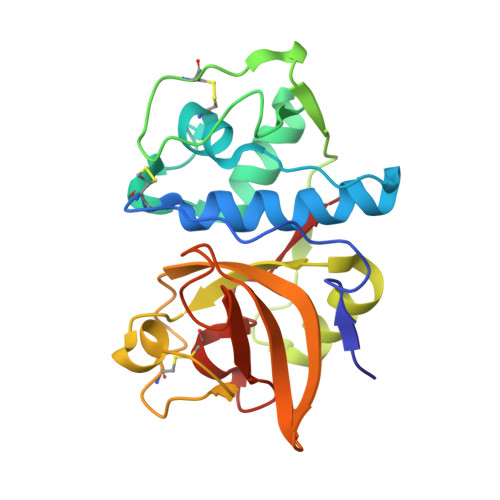
4N8W | pdb_00004n8w
cathepsin K - chondroitin sulfate complex
- PDB DOI: https://doi.org/10.2210/pdb4N8W/pdb
- Classification: HYDROLASE
- Organism(s): Homo sapiens
- Expression System: Komagataella pastoris
- Mutation(s): No
- Deposited: 2013-10-18 Released: 2014-11-26
- Deposition Author(s): Aguda, A.H., Nguyen, N.T., Bromme, D., Brayer, G.D.
Experimental Data Snapshot
- Method: X-RAY DIFFRACTION
- Resolution: 2.02 Å
- R-Value Free: 0.176 (Depositor), 0.181 (DCC)
- R-Value Work: 0.146 (Depositor), 0.152 (DCC)
- R-Value Observed: 0.149 (Depositor)
wwPDB Validation3D Report Full Report
Literature
- 👁 Image
Download Mendeley
Structural basis of collagen fiber degradation by cathepsin K.
Aguda, A.H., Panwar, P., Du, X., Nguyen, N.T., Brayer, G.D., Bromme, D.(2014) Proc Natl Acad Sci U S A 111: 17474-17479
- PubMed: 25422423 Search on PubMedSearch on PubMed Central
- DOI: https://doi.org/10.1073/pnas.1414126111
- Primary Citation Related Structures:
4N79, 4N8W - PubMed Abstract:
Cathepsin K is the major collagenolytic protease in bone that facilitates physiological as well as pathological bone degradation. Despite its key role in bone remodeling and for being a highly sought-after drug target for the treatment of osteoporosis, the mechanism of collagen fiber degradation by cathepsin K remained elusive. Here, we report the structure of a collagenolytically active cathepsin K protein dimer. Cathepsin K is organized into elongated C-shaped protease dimers that reveal a putative collagen-binding interface aided by glycosaminoglycans. Molecular modeling of collagen binding to the dimer indicates the participation of nonactive site amino acid residues, Q21 and Q92, in collagen unfolding. Mutations at these sites as well as perturbation of the dimer protein-protein interface completely inhibit cathepsin-K-mediated fiber degradation without affecting the hydrolysis of gelatin or synthetic peptide. Using scanning electron microscopy, we demonstrate the specific binding of cathepsin K at the edge of the fibrillar gap region of collagen fibers, which suggest initial cleavage events at the N- and C-terminal ends of tropocollagen molecules. Edman degradation analysis of collagen fiber degradation products revealed those initial cleavage sites. We propose that one cathepsin K molecule binds to collagen-bound glycosaminoglycans at the gap region and recruits a second protease molecule that provides an unfolding and cleavage mechanism for triple helical collagen. Removal of collagen-associated glycosaminoglycans prevents cathepsin K binding and subsequently fiber hydrolysis. Cathepsin K dimer and glycosaminoglycan binding sites represent novel targeting sites for the development of nonactive site-directed second-generation inhibitors of this important drug target.
- Department of Oral Biological and Medical Sciences, Faculty of Dentistry, Department of Biochemistry and Molecular Biology, Faculty of Medicine, and.
- Department of Biochemistry and Molecular Biology, Faculty of Medicine, and.
- Department of Oral Biological and Medical Sciences, Faculty of Dentistry, Department of Biochemistry and Molecular Biology, Faculty of Medicine, and Center for Blood Research, University of British Columbia, Vancouver, BC, Canada V6T 1Z3 dbromme@dentistry.ubc.ca.
Organizational Affiliation:
Explore in 3D: Structure | Sequence Annotations | Electron Density | Validation Report
Biological Assembly 1
Explore in 3D: Structure | Sequence Annotations | Electron Density | Validation Report
Global Symmetry: Asymmetric - C1
Global Stoichiometry: Monomer - A1
Find Similar Assemblies
Biological assembly 1 assigned by authors and generated by PISA (software)
Macromolecule Content
- Total Structure Weight: 24.92 kDa
- Atom Count: 1,807
- Modeled Residue Count: 213
- Deposited Residue Count: 215
- Unique protein chains: 1
Macromolecules
Entity ID: 1 | |||||
|---|---|---|---|---|---|
| Molecule | Chains | Sequence Length | Organism | Details | Image |
| Cathepsin K | 215 | Homo sapiens | Mutation(s): 0 Gene Names: CTSK, CTSO, CTSO2 EC: 3.4.22.38 | 👁 Image | |
UniProt & NIH Common Fund Data Resources | |||||
PHAROS: P43235 GTEx: ENSG00000143387 | |||||
Entity Groups | |||||
| Sequence Clusters | 30% Identity50% Identity70% Identity90% Identity95% Identity100% Identity | ||||
| UniProt Group | P43235 | ||||
Sequence AnnotationsExpand | |||||
Reference Sequence | |||||
Oligosaccharides
HelpEntity ID: 2 | |||||
|---|---|---|---|---|---|
| Molecule | Chains | Length | 2D Diagram | Glycosylation | D Interactions |
| 2-acetamido-2-deoxy-4-O-sulfo-beta-D-galactopyranose-(1-4)-beta-D-glucopyranuronic acid-(1-3)-2-acetamido-2-deoxy-4-O-sulfo-beta-D-galactopyranose-(1-4)-beta-D-glucopyranuronic acid-(1-3)-2-acetamido-2-deoxy-4-O-sulfo-beta-D-galactopyranose-(1-4)-beta-D-glucopyranuronic acid | B | 6 | 👁 Image | N/A | |
Glycosylation Resources | |||||
GlyTouCan: G16831RV GlyCosmos: G16831RV | |||||
Experimental Data & Validation
Experimental Data
- Method: X-RAY DIFFRACTION
- Resolution: 2.02 Å
- R-Value Free: 0.176 (Depositor), 0.181 (DCC)
- R-Value Work: 0.146 (Depositor), 0.152 (DCC)
- R-Value Observed: 0.149 (Depositor)
| Length ( Å ) | Angle ( ˚ ) |
|---|---|
| a = 31.717 | α = 90 |
| b = 67.504 | β = 90 |
| c = 92.869 | γ = 90 |
| Software Name | Purpose |
|---|---|
| ADSC | data collection |
| PHASES | phasing |
| REFMAC | refinement |
| MOSFLM | data reduction |
| SCALA | data scaling |
Entry History
Deposition Data
- Released Date: 2014-11-26 Deposition Author(s): Aguda, A.H., Nguyen, N.T., Bromme, D., Brayer, G.D.
Revision History (Full details and data files)
- Version 1.0: 2014-11-26
Type: Initial release - Version 1.1: 2014-12-24
Changes: Database references - Version 2.0: 2020-07-29
Type: Remediation
Reason: Carbohydrate remediation
Changes: Atomic model, Data collection, Derived calculations, Structure summary - Version 2.1: 2024-11-06
Changes: Data collection, Database references, Structure summary