
- FASTA Sequence
- PDBx/mmCIF Format
- PDBx/mmCIF Format (gz)
- BinaryCIF Format (gz)
- Legacy PDB Format
- Legacy PDB Format (gz)
- PDBML/XML Format (gz)
- Structure Factors (CIF)
- Structure Factors (CIF - gz)
- Validation Full (PDF - gz)
- Validation (XML - gz)
- Validation (CIF - gz)
- Validation 2fo-fc coefficients (CIF - gz)
- Validation fo-fc coefficients (CIF - gz)
- Biological Assembly 1 (CIF - gz)
- Biological Assembly 1 (PDB - gz)
4NEH | pdb_00004neh
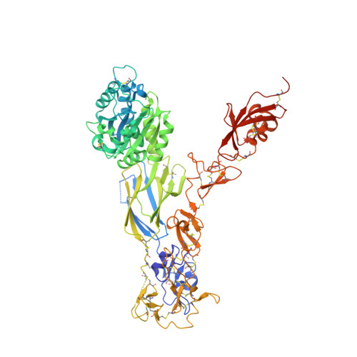
An internal ligand-bound, metastable state of a leukocyte integrin, aXb2
- PDB DOI: https://doi.org/10.2210/pdb4NEH/pdb
- Classification: CELL ADHESION
- Organism(s): Homo sapiens
- Expression System: Homo sapiens
- Mutation(s): No
- Deposited: 2013-10-29 Released: 2014-01-15
- Deposition Author(s): Sen, M., Yuki, K., Springer, T.A.
Experimental Data Snapshot
- Method: X-RAY DIFFRACTION
- Resolution: 2.75 Å
- R-Value Free: 0.225 (Depositor), 0.229 (DCC)
- R-Value Work: 0.193 (Depositor), 0.196 (DCC)
- R-Value Observed: 0.194 (Depositor)
Starting Models: experimental
View more details
Literature
- 👁 Image
Download Mendeley
An internal ligand-bound, metastable state of a leukocyte integrin, alpha X beta 2.
Sen, M., Yuki, K., Springer, T.A.(2013) J Cell Biol 203: 629-642
- PubMed: 24385486 Search on PubMedSearch on PubMed Central
- DOI: https://doi.org/10.1083/jcb.201308083
- Primary Citation Related Structures:
4NEH, 4NEN - PubMed Abstract:
How is massive conformational change in integrins achieved on a rapid timescale? We report crystal structures of a metastable, putative transition state of integrin αXβ2. The αXβ2 ectodomain is bent; however, a lattice contact stabilizes its ligand-binding αI domain in a high affinity, open conformation. Much of the αI α7 helix unwinds, loses contact with the αI domain, and reshapes to form an internal ligand that binds to the interface between the β propeller and βI domains. Lift-off of the αI domain above this platform enables a range of extensional and rotational motions without precedent in allosteric machines. Movements of secondary structure elements in the β2 βI domain occur in an order different than in β3 integrins, showing that integrin β subunits can be specialized to assume different intermediate states between closed and open. Mutations demonstrate that the structure trapped here is metastable and can enable rapid equilibration between bent and extended-open integrin conformations and up-regulation of leukocyte adhesiveness.
- Program in Cellular and Molecular Medicine, 2 Department of Medicine, 3 Department of Anethesiology, 4 Children's Hospital Boston, and 5 Department of Biological Chemistry and Molecular Pharmacology, Harvard Medical School, Boston, MA 02115.
Organizational Affiliation:
Explore in 3D: Structure | Sequence Annotations | Electron Density | Validation Report | Ligand Interaction (NAG)
Biological Assembly 1
Explore in 3D: Structure | Sequence Annotations | Electron Density | Validation Report | Ligand Interaction (NAG)
Global Symmetry: Asymmetric - C1
Global Stoichiometry: Hetero 2-mer - A1B1
Find Similar Assemblies
Biological assembly 1 assigned by authors and generated by PISA (software)
Macromolecule Content
- Total Structure Weight: 200.25 kDa
- Atom Count: 14,175
- Modeled Residue Count: 1,752
- Deposited Residue Count: 1,780
- Unique protein chains: 2
Macromolecules
Entity ID: 1 | |||||
|---|---|---|---|---|---|
| Molecule | Chains | Sequence Length | Organism | Details | Image |
| Integrin alpha-X | 1,094 | Homo sapiens | Mutation(s): 0 Gene Names: ITGAX, CD11C | 👁 Image | |
UniProt & NIH Common Fund Data Resources | |||||
PHAROS: P20702 GTEx: ENSG00000140678 | |||||
Entity Groups | |||||
| Sequence Clusters | 30% Identity50% Identity70% Identity90% Identity95% Identity100% Identity | ||||
| UniProt Group | P20702 | ||||
Glycosylation | |||||
| Glycosylation Sites: 5 | Go to GlyGen: P20702-1 | ||||
Sequence AnnotationsExpand | |||||
Reference Sequence | |||||
Entity ID: 2 | |||||
|---|---|---|---|---|---|
| Molecule | Chains | Sequence Length | Organism | Details | Image |
| Integrin beta-2 | 686 | Homo sapiens | Mutation(s): 0 Gene Names: ITGB2, CD18, MFI7 | 👁 Image | |
UniProt & NIH Common Fund Data Resources | |||||
PHAROS: P05107 GTEx: ENSG00000160255 | |||||
Entity Groups | |||||
| Sequence Clusters | 30% Identity50% Identity70% Identity90% Identity95% Identity100% Identity | ||||
| UniProt Group | P05107 | ||||
Glycosylation | |||||
| Glycosylation Sites: 2 | Go to GlyGen: P05107-1 | ||||
Sequence AnnotationsExpand | |||||
Reference Sequence | |||||
Oligosaccharides
HelpEntity ID: 3 | |||||
|---|---|---|---|---|---|
| Molecule | Chains | Length | 2D Diagram | Glycosylation | D Interactions |
| 2-acetamido-2-deoxy-beta-D-glucopyranose-(1-4)-2-acetamido-2-deoxy-beta-D-glucopyranose | C, F, G, H | 2 | 👁 Image | N-Glycosylation | |
Glycosylation Resources | |||||
GlyTouCan: G42666HT GlyCosmos: G42666HT GlyGen: G42666HT | |||||
Entity ID: 4 | |||||
|---|---|---|---|---|---|
| Molecule | Chains | Length | 2D Diagram | Glycosylation | D Interactions |
| beta-D-mannopyranose-(1-4)-2-acetamido-2-deoxy-beta-D-glucopyranose-(1-4)-2-acetamido-2-deoxy-beta-D-glucopyranose | D | 3 | 👁 Image | N-Glycosylation | |
Glycosylation Resources | |||||
GlyTouCan: G15407YE GlyCosmos: G15407YE GlyGen: G15407YE | |||||
Entity ID: 5 | |||||
|---|---|---|---|---|---|
| Molecule | Chains | Length | 2D Diagram | Glycosylation | D Interactions |
| alpha-D-mannopyranose-(1-2)-alpha-D-mannopyranose-(1-3)-[alpha-D-mannopyranose-(1-3)-alpha-D-mannopyranose-(1-6)]beta-D-mannopyranose-(1-4)-2-acetamido-2-deoxy-beta-D-glucopyranose-(1-4)-2-acetamido-2-deoxy-beta-D-glucopyranose | E | 7 | 👁 Image | N-Glycosylation | |
Glycosylation Resources | |||||
GlyTouCan: G07617FP GlyCosmos: G07617FP GlyGen: G07617FP | |||||
Small Molecules
Experimental Data & Validation
Experimental Data
- Method: X-RAY DIFFRACTION
- Resolution: 2.75 Å
- R-Value Free: 0.225 (Depositor), 0.229 (DCC)
- R-Value Work: 0.193 (Depositor), 0.196 (DCC)
- R-Value Observed: 0.194 (Depositor)
| Length ( Å ) | Angle ( ˚ ) |
|---|---|
| a = 126.967 | α = 90 |
| b = 131.444 | β = 90 |
| c = 190.477 | γ = 90 |
| Software Name | Purpose |
|---|---|
| PHASER | phasing |
| PHENIX | refinement |
| XDS | data reduction |
| XDS | data scaling |
Entry History
Deposition Data
- Released Date: 2014-01-15 Deposition Author(s): Sen, M., Yuki, K., Springer, T.A.
Revision History (Full details and data files)
- Version 1.0: 2014-01-15
Type: Initial release - Version 2.0: 2020-06-03
Changes: Atomic model, Data collection, Database references, Derived calculations - Version 3.0: 2020-07-29
Type: Remediation
Reason: Carbohydrate remediation
Changes: Advisory, Atomic model, Data collection, Derived calculations, Structure summary - Version 3.1: 2021-06-02
Changes: Source and taxonomy, Structure summary - Version 3.2: 2023-09-20
Changes: Data collection, Database references, Refinement description - Version 3.3: 2024-10-30
Changes: Structure summary